Clinical Anatomy Education in 2026: Integrating Dissection, Imaging, Simulation and Virtual Reality
Clinical anatomy education is entering a period of thoughtful transformation. Medical colleges are no longer limited to choosing between traditional dissection and digital learning. In 2026, the more useful question is how different educational methods can be combined to help students understand the human body accurately, retain anatomical knowledge and apply it safely in clinical practice.
Cadaveric dissection remains valuable for understanding natural variation, tissue layers, spatial relationships and professional responsibility. Medical imaging allows learners to study anatomy in living patients. Simulation provides a controlled environment for procedural practice. Three-dimensional models and virtual reality enable repeated exploration of complex structures from otherwise difficult viewpoints.
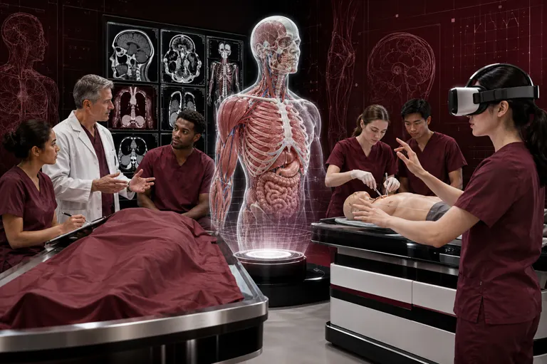
Each method addresses a different educational need. None provides a complete solution on its own.
An effective modern curriculum should connect structural knowledge with clinical examination, radiological interpretation, surgery, anaesthesia, pathology and patient safety. This requires more than adding a virtual-reality headset to an unchanged course. Learning objectives, teaching methods, assessments and faculty development must be aligned.
For the International Journal for Clinical Anatomy and Research, this evolution creates important opportunities for scholarly work in gross anatomy, radiological anatomy, surgical training, anatomical variation and medical education.
Why Clinical Anatomy Education Is Changing
Medical curricula face increasing pressure to cover more scientific and clinical material within limited teaching time. At the same time, students are expected to develop stronger diagnostic reasoning, imaging literacy and procedural awareness earlier in their education.
Anatomy departments are responding to several changes:
- Reduced time allocated to traditional anatomy courses
- Greater use of integrated and competency-based curricula
- Expansion of CT, MRI and ultrasonography in clinical care
- Increased availability of three-dimensional digital resources
- Growth of simulation-based medical education
- Wider use of virtual and augmented reality
- Greater attention to student-centred learning
- Need for repeatable and accessible teaching resources
- Increased emphasis on clinical application
- Recognition of anatomical variation and population diversity
These changes do not make foundational anatomy less important. They make the method of teaching it more consequential.
Students must move beyond memorising isolated structures. They need to understand where a structure is located, how it relates to neighbouring tissues, how it appears on imaging, how it may vary and why it matters in a clinical situation.
The Continuing Role of Cadaveric Dissection
Cadaveric dissection remains one of the most distinctive experiences in medical education. It allows learners to examine real human structure directly and to understand that anatomy is not perfectly uniform.
Three-Dimensional Spatial Understanding
Dissection requires students to work through layers. They see that a deep structure cannot be understood independently of the skin, fascia, muscles, vessels and nerves encountered before reaching it.
This layered learning helps students understand:
- Structural depth
- Organ relationships
- Tissue planes
- Neurovascular pathways
- Fascial compartments
- Surface landmarks
- Surgical approaches
- Routes through which disease may spread
The American Association for Anatomy notes that cadaveric study provides experience of tissue size, texture, depth and anatomical variation that virtual systems may not reproduce fully. American Association for Anatomy
Encountering Anatomical Variation
Textbooks usually show the most common pattern. Donors reveal variation in nerves, vessels, muscles, organs and skeletal structures.
This helps learners recognise that real patients may differ from diagrams. It also supports clinical reasoning by encouraging students to question unexpected findings rather than assume that the anatomy is incorrect.
Learning Through Exploration
Dissection is not entirely predictable. Students must locate structures, follow relationships and solve spatial problems. This investigative process can make learning more active and memorable.
Professional and Ethical Development
Working with donated human bodies introduces important lessons in dignity, confidentiality, death, gratitude and professional behaviour.
Institutions should prepare students for the emotional and ethical dimensions of the anatomy laboratory. Donors should be treated respectfully and used only within authorised educational and research purposes.
Limitations of Dissection
Cadaveric dissection also presents challenges:
- Limited availability of donated bodies
- Cost of laboratory infrastructure
- Preservation-related changes
- Chemical exposure
- Restricted teaching time
- Variation in student dissection quality
- Difficulty repeating a completed dissection
- Limited availability for remote learning
These limitations support integration with other methods rather than abandonment of cadaveric study.
Prosection as a Focused Teaching Method
Prosections are specimens prepared in advance to display specific anatomical relationships clearly.
They can be valuable when:
- Teaching time is limited
- Structures are difficult to expose
- A high-quality example should be shown to every student
- Clinical relationships need focused demonstration
- Learners require revision before assessment
Prosections can provide consistent teaching material, but students may lose some of the problem-solving experience involved in discovering structures through dissection.
A balanced curriculum may use dissection for exploration and prosections for reinforcement.
Integrating Medical Imaging into Anatomy Education
Medical imaging connects anatomy with living clinical practice. Students who learn structures only through diagrams and dissection may find it difficult to identify the same anatomy on a CT scan, MRI examination or ultrasound image.
Conventional Radiography
Radiographs introduce students to projectional anatomy. Learners must understand that multiple three-dimensional structures are superimposed in a two-dimensional image.
Radiography can support teaching of:
- Skeletal anatomy
- Joint alignment
- Chest anatomy
- Dental anatomy
- Basic pathological changes
- Surface and internal relationships
Computed Tomography
CT is particularly useful for teaching cross-sectional anatomy. Students can follow organs, vessels and tissue compartments through consecutive slices.
CT teaching may include:
- Axial, coronal and sagittal planes
- Thoracic anatomy
- Abdominal and pelvic organs
- Cranial structures
- Trauma anatomy
- Vascular anatomy
- Bone windows
- Soft-tissue windows
- Three-dimensional reconstruction
Magnetic Resonance Imaging
MRI offers detailed visualisation of soft tissues and is useful for teaching:
- Brain anatomy
- Spinal cord and nerves
- Musculoskeletal structures
- Pelvic anatomy
- Ligaments and tendons
- Joint compartments
- Organ relationships
Learners should understand that structures can appear differently across MRI sequences.
Ultrasonography
Ultrasound connects surface anatomy with real-time internal anatomy. Students must relate probe position to the image on the screen.
It can support teaching of:
- Vessels
- Peripheral nerves
- Muscles and tendons
- Abdominal organs
- Obstetric anatomy
- Cardiac anatomy
- Procedural landmarks
Ultrasound is particularly valuable for integrating anatomy with vascular access, regional anaesthesia and bedside examination.
Why Imaging Should Not Be Added in Isolation
Showing medical images without structured teaching may overwhelm beginners. Imaging should be introduced alongside the related anatomical region, relevant surface landmarks and clinical cases.
A useful sequence may involve:
- Study the region in an atlas
- Examine it through dissection or prosection
- Identify it on CT, MRI or ultrasound
- Apply the anatomy to a clinical case
- Review relevant anatomical variations
- Practise interpretation or a procedure
This creates connections across different forms of representation.
Simulation-Based Anatomy Education
Simulation allows learners to practise clinical tasks in a controlled environment before working with patients.
Procedural Simulation
Anatomical knowledge can be integrated into simulations involving:
- Airway management
- Vascular access
- Lumbar puncture
- Chest-drain insertion
- Regional nerve blocks
- Joint injection
- Endoscopy
- Laparoscopic procedures
- Fracture fixation
- Emergency interventions
Instead of memorising landmarks abstractly, students learn how anatomy guides an action.
Standardised and Repeatable Practice
A simulation can be repeated until the learner achieves a defined level of competence. Errors can be discussed without causing patient harm.
Feedback
Simulation is most effective when it includes structured feedback. Faculty can assess:
- Landmark identification
- Instrument position
- Procedural sequence
- Respect for vulnerable structures
- Communication
- Response to complications
Limitations
Mannequins and task trainers may simplify anatomy. They may not reproduce variation, tissue resistance or unexpected findings accurately.
Simulation should therefore be connected with real imaging, cadaveric material or clinically validated anatomical models.
Three-Dimensional Anatomical Models
Three-dimensional models allow learners to rotate, isolate, enlarge and reconstruct structures.
Digital 3D Models
Digital models can help students:
- View structures from multiple angles
- Remove superficial layers
- Isolate nerves or vessels
- Repeat difficult demonstrations
- Study outside the laboratory
- Explore relationships interactively
A 2025 study of a high-fidelity virtual dissection environment reported that interactive three-dimensional visualisation could provide enhanced learning experiences. PubMed study
Physical 3D Printing
Three-dimensional printing can convert imaging or anatomical datasets into physical models.
Potential uses include:
- Complex bone anatomy
- Congenital abnormalities
- Vascular pathways
- Surgical planning
- Skull-base relationships
- Implant teaching
- Patient-specific anatomy
Model Accuracy
A visually impressive model is not necessarily anatomically reliable. Institutions should evaluate:
- Source data
- Segmentation quality
- Resolution
- Anatomical validation
- Representation of small structures
- Presence of variations
- Intended educational level
Simplification may be useful for introductory teaching, but it should be disclosed.
Virtual Reality in Anatomy Education
Virtual reality places learners inside an interactive three-dimensional environment. Students may manipulate structures, change viewpoints and explore spaces that are difficult to appreciate through flat images.
Potential Educational Benefits
VR can support:
- Spatial understanding
- Learner engagement
- Repeated practice
- Self-directed learning
- Visualisation of deep structures
- Surgical orientation
- Collaborative learning
- Remote access
- Exploration of complex anatomical regions
Current Evidence in 2026
Research published in 2025 reported high student engagement with VR-based anatomy learning, with three-dimensional anatomical structures identified as a particularly valued feature. The study also identified the need for better onboarding, deeper content and stronger curriculum alignment. PubMed study
A prospective 2026 study found that one immersive VR session improved short-term anatomy knowledge and learner confidence, but the improvement was not sustained at delayed follow-up. The authors concluded that VR may be most useful as one part of a multimodal curriculum rather than as a standalone replacement. PubMed study
This distinction is important. High engagement does not automatically mean long-term learning.
Limitations of VR
Potential challenges include:
- Cost of hardware and licences
- Motion discomfort
- Limited device availability
- Technical support requirements
- Infection-control needs for shared headsets
- Accessibility concerns
- Variable digital literacy
- Cognitive overload
- Simplified anatomy
- Weak long-term retention after isolated sessions
The value of VR depends on instructional design, not merely the presence of technology.
Augmented Reality in Clinical Anatomy
Augmented reality overlays digital information onto a physical environment. It can place labels, structures or imaging data over a model, task trainer, donor or patient.
Potential applications include:
- Surgical anatomy teaching
- Image-guided procedures
- Surface-anatomy learning
- Regional anaesthesia
- Operative simulation
- Three-dimensional landmark identification
AR may help learners connect internal structures with external orientation, but registration accuracy is essential. An incorrectly aligned overlay can teach an incorrect relationship.
Photogrammetry and Digital Cadavers
Photogrammetry creates three-dimensional models from multiple photographs of a physical specimen.
This method can preserve valuable dissections digitally and provide learners with controlled access to rare or complex anatomical material.
A 2026 skull-base study produced photorealistic 360-degree models demonstrating cranial nerves, vessels and deep anatomical spaces. The authors presented the models as a complement to traditional dissection and conventional anatomical resources. PubMed study
Advantages
- Preservation of rare specimens
- Remote access
- Multiple viewing angles
- Repeatable demonstrations
- Integration with VR
- Reduced handling of delicate material
Ethical Responsibilities
Digital cadavers require the same respect as physical donors. Institutions should confirm that consent permits photography, scanning and digital distribution.
Donor identity must be protected, and access should be governed according to institutional policy.
Designing an Integrated Anatomy Curriculum
Adding more technologies does not automatically improve education. A curriculum needs a deliberate structure.
Start with Learning Outcomes
Every educational activity should address a defined outcome.
Examples include:
- Identify a structure
- Explain its relationships
- Recognise it on imaging
- Predict symptoms after injury
- Identify relevant variations
- Apply landmarks to a procedure
- Plan a surgical approach
The teaching method should be chosen according to the outcome.
Match the Method to the Learning Need
| Learning need | Suitable methods |
|---|---|
| Tissue layers and natural variation | Dissection and prosection |
| Cross-sectional recognition | CT and MRI |
| Surface-to-internal relationships | Ultrasound and AR |
| Repeated procedural practice | Simulation |
| Complex spatial orientation | 3D models and VR |
| Surgical corridors | Cadaveric training, imaging and simulation |
| Histological structure | Microscopy and virtual slides |
| Developmental processes | Models, animation and embryological imaging |
Use a Multimodal Sequence
A regional teaching unit may proceed through:
- Foundational terminology
- Atlas or three-dimensional model
- Dissection or prosection
- Radiological anatomy
- Clinical case
- Simulation or procedural application
- Virtual review
- Formative assessment
- Reflective or peer discussion
This sequence reinforces the same anatomy through several contexts.
Retrieval Practice and Spaced Learning
Anatomy is information-dense. One exposure is rarely sufficient for long-term retention.
Retrieval Practice
Instead of repeatedly rereading notes, learners should actively recall structures, relationships and clinical implications.
Retrieval activities may include:
- Spotter questions
- Image labelling
- Oral identification
- Short clinical cases
- Peer teaching
- Blank diagrams
- Radiology quizzes
- Timed digital assessments
Spacing
Important concepts should return throughout the curriculum. Anatomy taught in the first year should reappear during surgery, radiology, medicine and clinical-skills training.
Interleaving
Students should sometimes compare related regions, modalities or clinical problems rather than studying every structure in isolation.
Technology can support this process, but curriculum design remains the determining factor.
Anatomy Assessment in 2026
Assessment influences how students learn. If examinations test only labels and definitions, students may focus on short-term memorisation.
A modern assessment programme should evaluate:
- Structural identification
- Three-dimensional relationships
- Imaging interpretation
- Anatomical variation
- Clinical application
- Procedural landmarks
- Ethical understanding
- Communication of anatomy
Practical Spotters
Cadaveric, prosection, model and imaging stations can assess recognition across different representations.
Case-Based Questions
Clinical cases can ask students to identify the structure involved, explain symptoms or predict procedural risks.
Simulation Assessment
Procedural stations can evaluate whether learners use anatomy safely during an intervention.
Digital Assessment
Digital tools can provide rapid feedback, adaptive questions and repeated low-stakes practice.
However, automated scoring should not be used when the task requires complex clinical explanation or professional judgement.
Faculty Development
Educational technology cannot succeed without faculty preparation.
Teachers may need support in:
- Using VR and AR systems
- Teaching radiological anatomy
- Creating three-dimensional models
- Facilitating simulation
- Designing digital assessments
- Evaluating technology evidence
- Managing accessibility
- Protecting donor-related digital content
- Interpreting learning analytics
Technology should support the educator’s expertise rather than bypass it.
Accessibility and Educational Equity
A modern anatomy curriculum must consider whether all students can participate.
Potential barriers include:
- Limited access to devices
- Unreliable internet
- Visual or hearing impairment
- Motion sensitivity
- Physical disability
- Colour-vision differences
- Language barriers
- Digital inexperience
- Financial limitations
Institutions should provide alternative formats and avoid designing essential learning around a tool that some students cannot use.
Ethical Use of AI in Anatomy Education
Artificial intelligence may support anatomy teaching through personalised questions, image segmentation, feedback and adaptive learning.
Potential applications include:
- Generating practice questions
- Identifying weak learning areas
- Segmenting imaging data
- Supporting virtual tutors
- Providing terminology assistance
- Creating adaptive revision plans
Risks
AI tools may:
- Generate anatomically incorrect content
- Invent references
- oversimplify variation
- reproduce bias
- misuse personal learning data
- create overconfidence
- process donor images without appropriate permission
AI-generated anatomical information should be checked by qualified educators before use.
Connecting Anatomy Education with Patient Safety
The purpose of clinical anatomy education is not only examination performance. It is safer clinical practice.
Learners should understand how anatomy supports:
- Accurate diagnosis
- Imaging interpretation
- Safe injections
- Vascular access
- Regional anaesthesia
- Surgical planning
- Recognition of complications
- Protection of nerves and vessels
- Patient communication
A student who can label a structure but cannot apply it to a clinical decision has not yet achieved functional anatomical competence.
Research Priorities for Anatomy Education
The expansion of educational technology creates a need for rigorous research.
Important questions include:
- Does the method improve long-term retention?
- Does it improve spatial understanding?
- Can students transfer learning to imaging?
- Does it improve procedural performance?
- Which learners benefit most?
- What is the cognitive workload?
- How accessible is the technology?
- Is the method cost-effective?
- Does it improve patient-relevant outcomes?
- How does it compare with existing teaching?
- What combination of methods works best?
Beyond Student Satisfaction
Learners may enjoy a technology without learning more effectively. Studies should therefore avoid relying only on satisfaction surveys.
Stronger outcome measures include:
- Pre-intervention and post-intervention knowledge
- Delayed retention
- Practical examination performance
- Imaging interpretation
- Procedural accuracy
- Error rates
- Transfer to clinical tasks
- Validated confidence measures
- Cognitive workload
- Cost and implementation data
Appropriate Comparison Groups
A study comparing VR with no teaching may overestimate the value of VR. A more useful design compares it with an established teaching method or evaluates its benefit when added to a standard curriculum.
Reporting Anatomy Education Research
Researchers should report:
- Educational setting
- Learner level
- Sample size
- Learning objectives
- Intervention duration
- Faculty involvement
- Technology and software
- Comparison method
- Assessment tools
- Validation of instruments
- Statistical analysis
- Ethical approval
- Consent
- Missing data
- Limitations
- Conflicts of interest
- Technology-provider involvement
If the platform developer funded or participated in the study, this relationship should be disclosed.
IJCAR’s Role in Anatomy Education Research
The International Journal for Clinical Anatomy and Research provides a relevant scholarly platform for research connecting anatomy education with clinical practice.
Potential submissions include:
- Cadaveric teaching studies
- Imaging-integrated anatomy education
- Virtual-reality interventions
- Simulation-based training
- Three-dimensional model evaluation
- Anatomy assessment research
- Curriculum integration
- Learner retention studies
- Faculty-development research
- Digital histology education
- Donor ethics and professionalism
- Systematic reviews of anatomy teaching
IJCAR’s focus on clinical, gross, developmental, histological and radiological anatomy allows medical educators to examine how different teaching approaches contribute to clinically useful anatomical knowledge.
Researchers should consult the journal’s latest author instructions and ethical policies before submission.
Frequently Asked Questions
Is cadaveric dissection still necessary in 2026?
Cadaveric dissection remains valuable for learning tissue layers, spatial relationships, anatomical variation and professional responsibility. Its role may differ among institutions, but digital tools do not reproduce every part of the experience.
Can virtual reality replace cadaveric dissection?
Current evidence supports VR as a useful complement rather than a universal replacement. It offers repeatability and spatial visualisation but may not provide realistic tissue characteristics or natural variation.
How does medical imaging improve anatomy education?
CT, MRI and ultrasound help students identify anatomy in living patients and connect structural knowledge with diagnosis, surgery and clinical procedures.
What is simulation-based anatomy education?
It uses mannequins, task trainers, virtual systems or other controlled environments to teach anatomy through procedures and clinical scenarios.
What is the difference between VR and AR?
VR places the learner inside a fully digital environment. AR overlays digital information onto a physical environment, model or patient.
Are three-dimensional models always anatomically accurate?
No. Their quality depends on the source data, segmentation, validation and level of simplification. Educators should evaluate a model before using it.
Does VR improve long-term anatomy retention?
Some studies show short-term knowledge and engagement benefits, but lasting retention may require repeated sessions, retrieval practice and integration with other methods.
How should institutions select anatomy technology?
They should begin with learning objectives and evaluate anatomical accuracy, accessibility, evidence, technical support, cost and curriculum alignment.
Can AI be used to teach anatomy?
AI may support practice questions, adaptive learning and imaging analysis, but faculty must verify its output and protect learner and donor data.
What anatomy education studies can be submitted to IJCAR?
Relevant work may include studies of dissection, imaging, simulation, VR, AR, 3D models, anatomy assessment, curriculum design and clinically oriented teaching.
Conclusion
Clinical anatomy education in 2026 is not defined by replacing traditional teaching with digital technology. It is defined by purposeful integration.
Cadaveric dissection provides real structural complexity, tactile understanding and exposure to variation. Medical imaging connects anatomy with living patients. Simulation allows learners to practise clinical actions safely. Three-dimensional models improve visualisation, while virtual and augmented reality enable repeated exploration from new perspectives.
Each method has strengths and limitations. The most effective curriculum is likely to be multimodal, clinically integrated and supported by retrieval practice, appropriate assessment and trained faculty.
Educational innovation should be judged by more than novelty or student enjoyment. It should improve anatomical understanding, long-term retention, clinical transfer and patient safety.
The International Journal for Clinical Anatomy and Research encourages rigorous scholarship examining how anatomy can be taught more effectively across medical, surgical and allied health education. Researchers and educators working in dissection, imaging, simulation and immersive technology are invited to review IJCAR’s Aims and Scope and consider submitting relevant work.
Medical institutions or academic societies planning a scholarly journal may explore ScholarJMS for journal websites and editorial workflows, OJSCloud for OJS support, migration and ISSN consulting, GetDOI for Crossref DOI guidance, and Scholar9 for transparent peer-review support. Contact +91 82003 85143 through WhatsApp or email inquiry@ojscloud.com.