
Anatomical Variations and Their Clinical Significance in Diagnosis, Surgery and Medical Imaging
The human body follows a broadly recognisable structural plan, but it is not anatomically identical in every individual. Arteries may arise from an unusual source, nerves may divide at different levels, muscles may possess additional heads, organs may occupy variant positions, and bones may contain accessory processes or foramina.
Many of these differences are harmless and remain undiscovered throughout life. Others become clinically important when they alter symptoms, resemble disease on medical imaging, complicate a surgical approach or increase the risk of injury during an intervention.
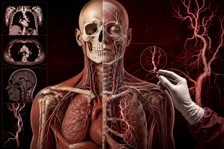
Anatomical variation is therefore not a minor exception to normal anatomy. It is an important part of human biological diversity and a central subject within clinical anatomy.
For clinicians, surgeons and radiologists, the key question is not simply whether a structure is different. The question is whether that difference changes diagnosis, procedural planning or patient management.
The International Journal for Clinical Anatomy and Research highlights the importance of studying anatomical variations through cadaveric dissection, radiological assessment, morphometric analysis, surgical observation and clinically meaningful case reporting. Such research connects foundational anatomical science with safer and more accurate healthcare.
What Is an Anatomical Variation?
An anatomical variation is a structural feature that differs from the pattern commonly described as typical but does not necessarily represent disease.
Variations may affect:
- The presence or absence of a structure
- The number of structures
- Size, shape or length
- Origin and insertion
- Branching pattern
- Position and orientation
- Course through the body
- Relationships with neighbouring tissues
- Fusion or separation of structures
- Vascular supply
- Nerve distribution
A variation may be congenital, developmental, population-related or associated with normal biological diversity.
Variation Is Not Always Abnormality
The terms “variation,” “anomaly” and “malformation” should not be used interchangeably without considering their meaning.
A normal anatomical variation is usually compatible with normal function and may have no clinical effect. A congenital anomaly may reflect an unusual developmental outcome that can have functional significance. A malformation generally implies a structural developmental defect with potential clinical consequences.
Accurate terminology matters because describing a harmless variant as abnormal may cause unnecessary concern or intervention.
The Concept of Normal Anatomy
Textbooks commonly present the most frequently observed pattern as standard anatomy. This creates an essential foundation for learning, but it does not represent every patient.
Normal anatomy is better understood as a range rather than one fixed configuration. Clinical competence requires knowledge of the expected pattern and awareness of the ways in which that pattern may vary.
How Anatomical Variations Develop
Many anatomical variations arise during embryonic development. Blood vessels, nerves, muscles and organs form through complex processes involving growth, migration, branching, fusion and regression.
A structure may vary when:
- A temporary embryonic vessel persists
- A vessel expected to remain disappears
- Tissue segments fuse incompletely
- A muscle separates into additional parts
- An organ follows a different rotational pathway
- A nerve develops an unusual connection
- One structure compensates for the absence of another
Genetic influences, developmental environment and population characteristics may all contribute. Not every variation has one clearly identifiable cause.
Understanding embryology can help explain why certain variations occur together and why some follow predictable patterns.
Why Anatomical Variations Matter in Clinical Diagnosis
Anatomical variation can change how symptoms appear, how physical findings are interpreted and which investigations are selected.
Variations May Alter Clinical Presentation
A variant nerve pathway may produce sensory or motor symptoms in a distribution that does not follow the usual textbook pattern. An accessory muscle may cause compression, swelling or exercise-related discomfort. An unusual vessel may contribute to pulsatile symptoms or affect blood flow.
If the clinician assumes that every patient follows standard anatomy, these presentations may be misunderstood.
Variations Can Resemble Disease
Some normal structures can appear mass-like, displaced or irregular. Without adequate anatomical knowledge, they may be mistaken for:
- Tumours
- Enlarged lymph nodes
- Vascular abnormalities
- Fractures
- Tendon injuries
- Organ enlargement
- Pathological calcification
- Developmental disease
This can lead to unnecessary investigations, referrals or interventions.
Variations Can Conceal Disease
A variant may also make genuine pathology harder to recognise. If an organ, vessel or duct lies in an unexpected location, a clinician may initially examine the wrong region or misinterpret the origin of symptoms.
Physical Examination May Be Affected
Surface landmarks are used to locate pulses, organs, bones, muscles and procedural sites. Variations in position or body structure can change the expected relationship between surface and internal anatomy.
Clinical examination should therefore be combined with imaging or other guidance when uncertainty could affect patient safety.
Vascular Variations and Their Clinical Importance
Variations in arteries and veins are among the most clinically significant anatomical differences because they can affect bleeding risk, organ perfusion, transplantation and image-guided treatment.
Arterial Branching Variations
Arteries may:
- Arise from an unusual parent vessel
- Divide earlier or later than expected
- Follow an atypical course
- Be duplicated
- Be absent
- Supply a different anatomical territory
- Persist from embryonic circulation
Such variations are important in abdominal, cardiovascular, transplant, neurological and reconstructive surgery.
Hepatic Arterial Variations
The liver may receive arterial supply through several different branching patterns. A replaced or accessory hepatic artery can affect liver transplantation, pancreatic surgery, hepatobiliary operations and embolisation procedures.
A multidetector CT angiography study involving 4,192 patients identified several replaced common hepatic artery patterns and emphasised their importance in upper abdominal surgical planning. PubMed study
The surgeon or interventional radiologist should understand not only that a variant exists but also which tissue depends on it for blood supply.
Renal Vascular Variations
Kidneys may receive blood through one or several renal arteries. Accessory arteries and early branching can influence donor nephrectomy, transplantation, renal surgery and endovascular procedures.
A 2025 CT angiography study of potential kidney donors reported renal arterial variations in a substantial proportion of the assessed participants, reinforcing the importance of preoperative vascular mapping. PubMed study
Venous Variations
Venous systems can also vary considerably. Duplicated veins, unusual drainage patterns, retroaortic vessels and persistent embryonic channels may complicate surgery or mimic pathology on imaging.
Unexpected venous injury can lead to major bleeding, particularly when the variant vessel is thin-walled or difficult to control.
Neural Variations in Clinical Practice
Peripheral nerves and central vascular-neural relationships frequently demonstrate variation.
Brachial Plexus Variations
The brachial plexus is a complex network supplying the upper limb. Variations may affect its roots, trunks, divisions, cords and terminal branches.
These differences can influence:
- Interpretation of nerve injury
- Regional anaesthesia
- Shoulder and axillary surgery
- Upper-limb reconstruction
- Nerve-transfer procedures
- Electrophysiological testing
IJCAR has published a detailed study of brachial plexus variations that discusses their relevance to nerve-injury diagnosis, regional blocks and surgical procedures involving the shoulder, axilla and upper arm. IJCAR brachial plexus study
Persistent Embryonic Vessels and Cranial Nerves
Persistent embryonic vessels may have close relationships with cranial nerves and the skull base. Their presence can alter blood flow and complicate neurosurgical or endovascular procedures.
An IJCAR cadaveric study described a persistent trigeminal artery connecting the internal carotid and basilar systems. The authors highlighted its possible relationship with trigeminal neuralgia and its significance during carotid, endovascular and skull-base procedures. IJCAR persistent trigeminal artery study
Nerve Branching and Communication
Nerves may communicate through unusual branches or divide at different levels. This can change patterns of weakness, sensory loss or neuropathic pain.
A clinician interpreting nerve-conduction findings or planning a nerve repair should consider the possibility that the patient’s neural anatomy differs from the most common pattern.
Musculoskeletal Variations
Musculoskeletal variations may involve bones, muscles, tendons, ligaments and joints.
Accessory Muscles and Tendons
Accessory or duplicated muscles may be asymptomatic. In some patients, however, they can cause nerve compression, vascular compression, pain or reduced movement.
On imaging, an unfamiliar muscle may be misinterpreted as a soft-tissue mass.
Bone and Foramen Variations
Accessory bones, unusual processes and variant foramina may resemble fractures or alter the route of nerves and vessels.
Correct identification is particularly important in trauma, orthopaedics, dentistry, spinal surgery and skull-base procedures.
Ligament Variations
Ligament anatomy influences joint stability, reconstruction and imaging interpretation. Variation in the presence, dimensions or attachment of a ligament may affect operative planning.
IJCAR has published a cadaveric study examining variations of the anterolateral ligament of the knee and their implications for reconstructive arthroscopic surgery. IJCAR anterolateral ligament study
This type of investigation demonstrates how detailed anatomical research can contribute directly to surgical decision-making.
Anatomical Variations in Medical Imaging
Medical imaging has made it possible to study anatomy in living patients across large and diverse populations. It also creates an important responsibility: the interpreter must distinguish normal variation from disease.
Conventional Radiography
Projectional radiographs compress three-dimensional structures into two-dimensional images. Overlapping tissues and variant positions can create misleading appearances.
Accessory bones, unusual joint contours and developmental differences may be mistaken for fractures or degenerative disease.
Computed Tomography
CT provides detailed cross-sectional and reconstructed images of bones, organs, vessels and tissue relationships.
It is particularly useful for:
- Mapping vascular variations
- Examining complex skeletal anatomy
- Assessing congenital differences
- Planning surgery
- Evaluating trauma
- Creating three-dimensional models
Contrast-enhanced CT and CT angiography can demonstrate arterial origin, branching, course and organ supply.
Magnetic Resonance Imaging
MRI provides high soft-tissue contrast and is valuable for evaluating:
- Brain and spinal structures
- Cranial and peripheral nerves
- Muscles
- Tendons
- Ligaments
- Joints
- Pelvic organs
- Vascular relationships
A variant muscle, nerve or vascular structure may be clinically important even when it is not pathological.
Ultrasonography
Ultrasound offers dynamic, real-time assessment but depends significantly on the operator’s anatomical understanding.
Variant anatomy may affect:
- Vascular access
- Nerve blocks
- Biopsy
- Fluid drainage
- Obstetric assessment
- Abdominal imaging
- Musculoskeletal procedures
The operator should explore structures systematically rather than relying on a single expected view.
Angiography
Angiographic methods are valuable for planning vascular and interventional procedures. They can show arterial and venous variants that may change catheter routes, embolisation strategies or surgical access.
Diagnostic Errors Associated with Anatomical Variations
Anatomical variants can contribute to several types of diagnostic error.
False-Positive Interpretation
A normal variant may be reported as disease. This can expose patients to repeat imaging, invasive tests or unnecessary treatment.
False-Negative Interpretation
A genuine abnormality may be overlooked when it occurs in an unexpected location or when variant anatomy changes the expected appearance.
Incorrect Attribution
A lesion may be assigned to the wrong organ or tissue because the underlying anatomy differs from expectation.
Incomplete Reporting
A radiologist may identify a variant but omit it from the report because it appears incidental. If the patient later undergoes surgery, the missing information may become highly relevant.
A clinically useful report should mention variations that could influence intervention, surgery or diagnosis, even when they are not the direct cause of the patient’s symptoms.
Anatomical Variations and Surgical Planning
Surgery is one of the settings in which a previously unknown variation can have immediate consequences.
Preoperative Evaluation
Before complex procedures, clinicians should review imaging for:
- Variant vessels
- Abnormal nerve courses
- Organ position
- Ductal anatomy
- Skeletal morphology
- Previous surgical changes
- Congenital differences
- Relationships between lesions and surrounding structures
A standard radiology report may not always address every relationship relevant to a specific operation. Direct discussion between the surgeon and radiologist can improve planning.
Intraoperative Recognition
Not every variation is detected before surgery. Surgeons must remain alert when anatomy does not match the expected pattern.
Safe responses include:
- Improving exposure
- Confirming landmarks
- Avoiding blind division
- Reviewing imaging
- Using intraoperative imaging where appropriate
- Seeking specialist assistance
- Modifying the surgical approach
Vascular Control
A variant artery should not be divided until its origin, course and territory are understood. An apparently small vessel may provide essential blood supply to an organ segment.
Nerve Preservation
Variant nerve branching can make standard danger zones less predictable. Identification based on several landmarks is safer than relying on one expected position.
Implant and Graft Placement
Differences in bone morphology, vessel supply, ligament attachment or soft-tissue dimensions may influence implant size, graft selection and fixation.
Anatomical Variations in Regional Anaesthesia and Clinical Procedures
Anaesthesiologists and procedural specialists frequently depend on anatomical landmarks.
Variations may influence:
- Peripheral nerve blocks
- Central venous access
- Arterial cannulation
- Lumbar puncture
- Epidural anaesthesia
- Chest drainage
- Joint injection
- Biopsy
- Catheter placement
- Endoscopic procedures
Regional Nerve Blocks
An altered nerve course or branching pattern may reduce block effectiveness or increase injury risk. Ultrasound guidance can help identify the target and nearby vessels, but the operator must still understand regional anatomy.
Vascular Access
Duplicated vessels, unusual courses and altered relationships may complicate catheter placement. Real-time imaging and careful route selection can improve safety.
Spinal Procedures
Variation in vertebral anatomy, segmentation and surface landmarks can affect identification of the correct level. Imaging confirmation may be necessary when the consequences of incorrect localisation are significant.
Anatomical Variations and Patient Safety
Anatomical knowledge contributes to patient safety by helping clinicians anticipate rather than merely react to unexpected structures.
Preventing Iatrogenic Injury
Unrecognised variants can contribute to:
- Haemorrhage
- Nerve damage
- Organ injury
- Ischaemia
- Failed procedures
- Incomplete surgery
- Incorrect level identification
- Postoperative dysfunction
Preoperative imaging, careful dissection and accurate communication reduce these risks.
Improving Team Communication
When a radiologist identifies a clinically important variation, the finding should be communicated clearly. The report should describe the structure, course and likely procedural relevance.
Surgeons should inform the operative team when a variant changes the planned approach.
Recording Variants for Future Care
Important anatomical findings should remain available in the patient’s medical record. A variation discovered during one procedure may affect future treatment.
The Role of Cadaveric Research
Cadaveric research remains essential for identifying and documenting variations that may not be clearly visible on routine imaging.
Advantages of Cadaveric Investigation
Dissection allows researchers to examine:
- Tissue layers
- Small branches
- Deep anatomical relationships
- Origins and insertions
- Communications between structures
- Measurements
- Bilateral differences
- Relationships with fixed landmarks
Cadaveric work can also test surgical approaches and identify vulnerable structures.
Importance of Methodological Detail
Researchers should report:
- Number of specimens
- Age and sex data where available
- Preservation method
- Inclusion and exclusion criteria
- Dissection approach
- Measurement instruments
- Observer reliability
- Photography or documentation method
- Ethical or institutional approval
- Study limitations
A case report may be valuable when a variation is rare or clinically important, but broader conclusions should not be drawn from one specimen.
Imaging-Based Variation Research
CT, MRI, ultrasonography and angiography allow researchers to study living anatomy across larger samples.
Imaging-based research can examine:
- Prevalence of variants
- Differences among populations
- Sex-related patterns
- Age-related changes
- Bilateral symmetry
- Morphometric reference ranges
- Surgical-risk relationships
- Correlation with symptoms
Population-Specific Research
Anatomical prevalence reported in one population may not apply identically to another. Well-designed regional studies can contribute valuable information, especially when sampling methods and imaging criteria are reported transparently.
However, researchers should avoid interpreting limited samples as proof of fixed racial or ethnic categories. Findings should be presented within the limits of the study population.
Three-Dimensional Models and Artificial Intelligence
New technologies are expanding the study and application of anatomical variation.
Three-Dimensional Reconstruction
Three-dimensional reconstructions can make complex vascular, skeletal and organ relationships easier to understand. They can support:
- Preoperative planning
- Surgical rehearsal
- Implant design
- Anatomy education
- Patient communication
- Morphometric research
Three-Dimensional Printing
Patient-specific models may help surgical teams compare approaches before complex procedures.
Artificial Intelligence
AI-assisted tools can segment structures, detect landmarks and measure anatomy on imaging data. These capabilities may make large-scale variation research more efficient.
However, AI models may perform less reliably when they encounter anatomy that was underrepresented in their training data. Human anatomical expertise remains necessary for recognising unusual patterns and verifying automated results.
Reporting an Anatomical Variation for Publication
A strong anatomical-variation manuscript should do more than describe an uncommon structure.
Authors should explain:
- What differs from the expected pattern
- How the variation was identified
- Whether it was unilateral or bilateral
- Relevant measurements
- Embryological explanation where appropriate
- Comparison with previous evidence
- Diagnostic significance
- Surgical or procedural implications
- Limitations of the observation
Use Clear Visual Documentation
High-quality images, diagrams and labels are essential. Readers should be able to understand the spatial relationships without relying only on written description.
Avoid Exaggerating Rarity
Authors should search the literature carefully before claiming that a variation is the first or rarest report. Similar findings may have been described using different terminology.
Explain Clinical Relevance Precisely
Statements such as “surgeons should be aware” are not sufficient on their own. Authors should state which procedure may be affected, which structure is at risk and how clinical practice may need to change.
IJCAR’s Role in Anatomical Variation Research
The International Journal for Clinical Anatomy and Research provides an appropriate platform for studies that connect anatomical variation with diagnosis, surgery, imaging and patient safety.
Relevant submissions may include:
- Cadaveric studies
- Imaging-based anatomical research
- Vascular variation studies
- Neural variation studies
- Musculoskeletal anatomy
- Morphometric investigations
- Anatomical case reports
- Surgical anatomy
- Radiological correlation
- Developmental anatomy
- Systematic reviews
- Three-dimensional anatomical modelling
IJCAR’s current content includes research on the anterolateral ligament of the knee, brachial plexus variations, nutrient artery foramina and persistent embryonic vessels. These studies reflect the journal’s role in connecting structural anatomy with clinical practice.
Researchers should review the journal’s aims, ethical policies and author guidelines before submitting their work.
Frequently Asked Questions
What is an anatomical variation?
An anatomical variation is a structural difference from the pattern commonly described as typical. It may involve the number, size, position, origin, course or relationship of a body structure.
Are anatomical variations diseases?
Most anatomical variations are not diseases. Many are harmless, but some become clinically significant during diagnosis, surgery or another procedure.
Why are anatomical variations important in surgery?
They may change the position of vessels, nerves, ducts, muscles or organs. If an important variation is not recognised, it can increase the risk of bleeding, nerve injury or organ damage.
Can anatomical variants resemble tumours?
Yes. Accessory muscles, unusual vessels, developmental remnants and variant organs may resemble masses or other pathological findings on imaging.
Which imaging method is best for detecting variations?
The appropriate modality depends on the structure. CT angiography is useful for many vascular patterns, MRI provides detailed soft-tissue and neural anatomy, and ultrasound offers real-time assessment.
How do vascular variations affect transplantation?
Accessory or replaced vessels can affect organ removal, reconstruction and blood supply. Preoperative vascular mapping is therefore important in transplant planning.
Can a normal variation cause symptoms?
Yes. A variation may compress a nerve or vessel, alter blood flow or change biomechanics. Many others remain completely asymptomatic.
Why is cadaveric research still important?
Cadaveric dissection reveals small branches, tissue planes and deep structural relationships that may not be fully visible on routine imaging.
Can artificial intelligence detect anatomical variations?
AI can assist with segmentation and landmark identification, but unusual anatomy may be underrepresented in training data. Expert verification remains necessary.
What anatomical-variation studies can be submitted to IJCAR?
IJCAR may consider cadaveric, radiological, morphometric, surgical, developmental and clinically relevant case studies that align with its current scope and author guidelines.
Conclusion
Anatomical variation is an expected part of human structural diversity. Its clinical importance depends on the structure involved, its relationship with surrounding tissues and the procedure or diagnosis under consideration.
For clinicians, recognising variations can improve diagnostic reasoning. For radiologists, it can prevent normal anatomy from being mistaken for disease. For surgeons and procedural specialists, it can reduce unexpected bleeding, nerve injury, organ damage and technical failure.
Cadaveric dissection, medical imaging, intraoperative observation and three-dimensional technologies each contribute different evidence. When these methods are combined, researchers can build a more complete understanding of human anatomical diversity.
The International Journal for Clinical Anatomy and Research encourages scientifically rigorous work that explains not only what anatomical variation exists but why it matters to clinical care. Research that connects anatomical observation with diagnosis, medical imaging, surgery and patient safety can make a meaningful contribution to both anatomical science and modern medicine.
Authors working in these areas are encouraged to consult IJCAR’s Aims and Scope, Author Guidelines and online manuscript-submission page.
Medical institutions or scholarly societies planning their own journal can explore ScholarJMS for journal websites and editorial workflows, OJSCloud for OJS support, migration and ISSN consulting, GetDOI for Crossref DOI workflow guidance, and Scholar9 for transparent peer-review support. Contact +91 82003 85143 through WhatsApp or email inquiry@ojscloud.com.