Surgical Anatomy and Patient Safety: Why Anatomical Precision Matters in Clinical Practice
Every surgical procedure is ultimately performed within a complex anatomical environment. Whether the intervention is a routine incision, a laparoscopic procedure, a vascular reconstruction or a highly specialised operation, its safety depends on the surgeon’s ability to identify structures, understand their relationships and anticipate how they may differ among individual patients.
Surgical anatomy is the application of anatomical knowledge to operative and procedural practice. It examines the structures encountered during surgery, the pathways used to reach them and the anatomical relationships that must be protected. It also considers how disease, trauma, development, ageing and previous treatment may alter normal anatomy.
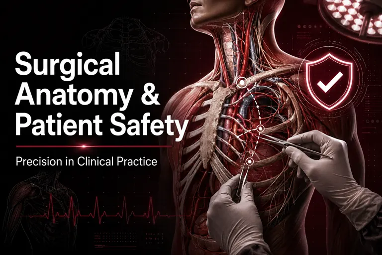
Advances in imaging, minimally invasive surgery, robotic systems, navigation and artificial intelligence have significantly changed modern surgical practice. Nevertheless, these tools do not reduce the need for anatomical knowledge. They require surgeons to apply that knowledge with even greater precision.
Anatomical accuracy supports correct diagnosis, appropriate surgical planning, safe tissue dissection, control of bleeding and preservation of neurological and organ function. When anatomy is misunderstood or an important variation is overlooked, the consequences may include haemorrhage, nerve injury, organ damage, incomplete treatment or avoidable postoperative complications.
For the International Journal for Clinical Anatomy and Research, the relationship between surgical anatomy and patient safety is a central area of scholarly importance. Research involving gross anatomy, radiological anatomy, morphometry, anatomical variations and operative approaches can help translate structural knowledge into safer clinical care.
What Is Surgical Anatomy?
Surgical anatomy focuses on the structures and relationships that matter during an operation or clinical procedure. It extends beyond the identification of organs, muscles, vessels and nerves. It examines how these structures are approached, exposed, separated, protected, repaired or removed.
A surgical understanding of anatomy includes:
- The arrangement of structures within a particular region
- The layers encountered from the surface to the operative target
- The course and branches of arteries and veins
- The position of nerves and the functions they control
- Fascial planes, tissue compartments and potential spaces
- The relationships between organs and surrounding structures
- Anatomical landmarks used for orientation
- Common and uncommon anatomical variations
- Changes caused by pathology, trauma or previous surgery
This knowledge allows surgeons to form a three-dimensional mental map of the operative region. It helps them anticipate what should appear at each stage and recognise when the anatomy differs from expectation.
Why Anatomical Precision Matters in Surgery
Anatomical precision means identifying and working with structures accurately, consistently and in relation to the individual patient. It is not limited to knowing where a structure is usually found. It includes recognising its boundaries, variations, depth, orientation and clinical importance.
A small anatomical error can have major consequences. A few millimetres may separate a safe dissection plane from an important nerve, vessel or duct. In confined regions such as the skull base, neck, pelvis, hand and spine, this margin may be particularly narrow.
Anatomical precision influences several dimensions of surgical quality:
- Selection of an appropriate operative approach
- Accurate identification of the target structure
- Protection of neighbouring tissues
- Control of vascular structures
- Preservation of neurological function
- Completeness of tumour removal
- Positioning of implants and grafts
- Reduction of operative time and tissue trauma
- Prevention of avoidable complications
Technical skill remains essential, but technique cannot be separated from anatomical understanding. Safe surgical movement depends on knowing which structure is being handled, what lies beside it and what may be encountered next.
Surgical Anatomy and Preoperative Planning
Patient safety begins before the first incision. Preoperative planning allows the surgical team to understand the clinical problem, review the patient’s anatomy and prepare for possible difficulties.
Defining the Operative Target
The surgical team must identify the precise structure or region requiring treatment. A lesion may appear to arise from one organ while actually originating from an adjacent compartment. A fracture may involve an articular surface, neurovascular canal or ligament attachment that changes the operative strategy.
Clear anatomical localisation helps ensure that the selected procedure addresses the correct pathology.
Reviewing Patient-Specific Anatomy
Textbooks describe common anatomical patterns, but every operation is performed on an individual patient. Preoperative imaging may reveal differences in organ position, vessel branching, skeletal morphology or the relationship between a lesion and surrounding structures.
Relevant imaging may include:
- Conventional radiography
- Ultrasonography
- Computed tomography
- Magnetic resonance imaging
- CT or MR angiography
- Three-dimensional reconstruction
- Functional or nuclear imaging
The surgeon should interpret these findings within the context of the planned procedure rather than relying only on a general imaging report.
Selecting the Surgical Approach
Several approaches may provide access to the same anatomical region. The most appropriate choice depends on the location of the target, the surrounding anatomy, the patient’s condition and the surgeon’s objectives.
An approach should provide sufficient exposure while limiting unnecessary disruption of normal tissue. It should also allow control of critical structures if unexpected bleeding or another complication occurs.
Anticipating Anatomical Difficulty
Disease can distort anatomy. Tumours may displace or invade tissues. Infection can eliminate normal planes. Trauma may fragment landmarks. Previous operations can cause adhesions and scar formation.
Recognising these possibilities before surgery allows the team to prepare additional equipment, seek specialist support and develop alternative strategies.
Anatomical Landmarks in Surgical Orientation
Anatomical landmarks help surgeons locate structures and maintain orientation. They may include bones, vessels, nerves, muscles, tendons, ducts, fascial boundaries or identifiable organ surfaces.
Reliable landmarks are especially important when the operative target is not immediately visible. A surgeon may follow one known structure to locate another or use a fixed bony point to estimate the course of a nerve or vessel.
However, landmarks must be used carefully. Their position may change because of:
- Patient age and body composition
- Congenital differences
- Disease-related displacement
- Trauma
- Previous surgery
- Surgical positioning
- Tissue traction or insufflation
A landmark is therefore a guide rather than an absolute guarantee. Safe surgery requires confirmation from multiple anatomical relationships whenever possible.
Tissue Planes and Safe Surgical Dissection
Many surgical procedures depend on identifying natural tissue planes. These planes allow tissues to be separated with less bleeding and reduced damage to surrounding structures.
Fascial Planes
Fascia surrounds muscles, organs, vessels and tissue compartments. Some fascial planes provide useful routes for surgical exposure. Others help contain or direct the spread of blood, infection or fluid.
Understanding fascial anatomy helps surgeons select appropriate dissection pathways and anticipate how disease may extend beyond its apparent origin.
Embryological Planes
Certain operative planes develop during embryological growth as organs rotate, fuse or become attached to surrounding tissues. These planes may contain relatively fewer vessels and can provide safe routes during abdominal or pelvic surgery.
Knowledge of developmental anatomy can therefore have direct operative value.
Sharp and Blunt Dissection
The choice between sharp and blunt dissection depends on tissue characteristics and anatomical relationships. Blunt dissection may be useful in loose planes, while sharp dissection may be required near delicate or adherent structures.
Neither method is automatically safe. The surgeon must understand what lies beyond the visible field and how much force the tissues can tolerate.
Anatomical Variations and Surgical Risk
Anatomical variation is one of the most important reasons why a standard procedure cannot be performed mechanically. Structures may differ in number, origin, course, size, branching pattern or relationship to surrounding tissues.
Vascular Variations
Arterial and venous patterns frequently vary. An accessory artery, unusual branching pattern or altered venous drainage may influence surgical access and bleeding risk.
Vascular variations are particularly important in:
- Transplantation
- Hepatobiliary surgery
- Vascular surgery
- Neurosurgery
- Head and neck surgery
- Renal surgery
- Orthopaedic reconstruction
- Flap and graft procedures
Preoperative angiographic imaging can identify many variations, but the surgical team must remain prepared for findings that were not recognised beforehand.
Neural Variations
Nerves may divide earlier or later than expected, follow an unusual course or have variable relationships with nearby structures. Injury can result in sensory loss, weakness, pain or dysfunction of an organ.
Detailed knowledge of neural anatomy is particularly important in surgery involving the neck, upper limb, pelvis, spine and cranial base.
Musculoskeletal Variations
Accessory muscles, variant tendons, altered ligament attachments and differences in bone morphology can affect exposure, reconstruction and implant placement. Some variations may also be mistaken for pathological tissue or previous injury.
Visceral and Ductal Variations
Variations in biliary, pancreatic, renal, pulmonary and gastrointestinal anatomy may change the safest operative approach. Failing to recognise them can lead to incomplete treatment or injury to an unexpected duct, vessel or organ segment.
Why Variation Research Matters
Cadaveric studies, radiological investigations and surgical case reports help document the range of human anatomy. Their value increases when authors explain how a variation affects clinical decisions.
An anatomical variation should not merely be described as rare. Researchers should clarify:
- How it can be recognised
- Whether it can be detected before surgery
- Which procedures it may affect
- What complications could occur
- How the surgical technique may be modified
This clinical interpretation transforms an anatomical observation into actionable patient-safety knowledge.
Protecting Blood Vessels During Surgery
Vascular injury can cause rapid blood loss, reduced organ perfusion, haematoma formation or the need for emergency conversion to a more extensive procedure.
Safe vascular management begins with understanding the expected location and branching pattern of major vessels. It also requires awareness of smaller branches that may be clinically important.
Important principles include:
- Identifying vessels before division or ligation
- Obtaining adequate exposure
- Avoiding blind clamping or cutting
- Recognising vascular variants
- Maintaining control of proximal and distal segments
- Confirming perfusion after reconstruction
- Understanding collateral circulation
Preoperative vascular imaging may provide valuable information, but intraoperative recognition remains essential.
Preventing Nerve Injury
Nerves may be difficult to identify because they are small, embedded in tissue or displaced by disease. Their injury can produce temporary or permanent functional loss.
Direct and Indirect Nerve Injury
A nerve may be damaged by cutting, stretching, compression, heat, ischaemia or entrapment within a suture or implant. Protection therefore requires more than avoiding direct transection.
The surgical team should understand:
- The expected course of the nerve
- Its relationship to operative landmarks
- Possible branching variations
- Areas where it is fixed and vulnerable to traction
- The function that may be lost if it is injured
In selected procedures, nerve monitoring may support identification and functional preservation. It complements but does not replace anatomical knowledge.
Surgical Anatomy in Minimally Invasive Procedures
Laparoscopic, arthroscopic and endoscopic procedures reduce the size of external incisions but can make anatomical orientation more challenging.
Restricted Field of View
The camera shows only part of the operative field. Structures outside the visible area may still be vulnerable to injury. The surgeon must maintain awareness of the wider anatomical environment.
Loss of Direct Tactile Feedback
In conventional surgery, palpation helps identify tissue consistency, vessel pulsation and lesion boundaries. Minimally invasive procedures may limit this feedback, increasing dependence on visual anatomy and preoperative imaging.
Altered Perspective
The magnified camera view may approach tissues from angles that differ from conventional dissection. Familiar structures can appear unfamiliar when rotated or viewed from within a cavity.
Instrument Access
Instruments enter through fixed access points. Their angle, depth and range of movement must be planned in relation to the target and surrounding structures.
Safe minimally invasive surgery therefore depends on strong three-dimensional understanding and the ability to interpret anatomy from changing viewpoints.
Surgical Anatomy in Robotic Surgery
Robotic platforms can provide magnified three-dimensional visualisation, stable instruments and precise movement. These advantages can support detailed dissection in confined spaces.
However, robotic surgery introduces additional anatomical considerations:
- The surgeon may be physically separated from the patient
- Instrument movement is mediated through a console
- Tactile feedback may be limited
- Visual magnification can reduce awareness of the wider field
- Safe port placement requires surface and internal anatomy
- Emergency access must be considered before docking
Robotic precision cannot compensate for incorrect anatomical interpretation. The technology performs the surgeon’s intended movement, so the safety of that movement still depends on knowledge and judgement.
Surgical Anatomy and Image-Guided Procedures
Image-guided surgery combines preoperative or intraoperative imaging with navigation technology. It is used in neurosurgery, spinal surgery, orthopaedics, otolaryngology and other specialties.
Navigation can help localise a target, identify instrument position and guide treatment near important structures. Three-dimensional models can also help teams rehearse complex procedures using patient-specific anatomy.
The American College of Surgeons has highlighted the value of patient-specific modelling and three-dimensional printing in preoperative planning, particularly when teams need to compare possible approaches before surgery. American College of Surgeons
Navigation systems nevertheless have limitations. Registration errors, patient movement and changes in tissue position during surgery can reduce accuracy. Surgeons must verify that digital guidance corresponds with actual anatomy.
Wrong-Site and Wrong-Level Surgery
Anatomical precision also includes confirming the correct patient, procedure, side and anatomical level.
Wrong-site, wrong-procedure and wrong-patient operations are considered serious preventable safety events. They may involve the wrong limb, organ, side or spinal level. Prevention requires structured verification rather than reliance on memory or assumption. AHRQ Patient Safety Network
Important measures include:
- Confirming patient identity
- Reviewing consent and clinical documentation
- Matching imaging with the planned procedure
- Marking the operative site when appropriate
- Verifying laterality
- Confirming implants and equipment
- Conducting a team time-out
- Using intraoperative imaging when anatomical level is uncertain
The World Health Organization Surgical Safety Checklist was developed to reduce errors, adverse events and communication failures in surgery. It promotes team confirmation of essential safety information before anaesthesia, incision and departure from the operating room. World Health Organization
A checklist cannot replace anatomy, but it creates an organised opportunity to verify that anatomical and procedural decisions are correct.
The Relationship Between Anatomy and Iatrogenic Injury
Iatrogenic injury is harm unintentionally caused during medical treatment. Some injuries occur because a normal structure lies close to the operative pathway. Others result from unusual anatomy, poor exposure or disease-related distortion.
Common anatomy-related risks include:
- Nerve damage
- Vascular injury
- Organ perforation
- Ductal injury
- Damage to adjacent muscles or tendons
- Incorrect implant positioning
- Injury caused by excessive instrument depth
- Compartment or fascial-plane violation
Understanding the mechanism of these injuries is important for both prevention and education. Detailed complication reports can help other clinicians recognise dangerous anatomical situations and improve operative techniques.
Surgical Anatomy in Emergency and Trauma Care
Emergency surgery presents special anatomical challenges. Decisions may need to be made quickly, and normal anatomy may be severely disrupted.
Trauma-Related Distortion
Fractures, bleeding, swelling and tissue disruption can obscure landmarks. The operative team must reconstruct the likely anatomy from imaging, clinical examination and direct exploration.
Haemorrhage Control
Rapid control of bleeding depends on identifying the injured vessel or the anatomical region from which it arises. Knowledge of vascular territories and collateral pathways can guide temporary and definitive control.
Damage-Control Surgery
In unstable patients, the immediate objective may be to control haemorrhage and contamination rather than complete reconstruction. Anatomical knowledge helps the surgeon prioritise structures and select the shortest safe route to achieve those goals.
Surgical Anatomy Education and Training
Anatomical competence develops through repeated learning and clinical application. Textbooks provide a foundation, but surgical understanding also requires three-dimensional exposure and procedural context.
Cadaveric Dissection
Cadaveric dissection remains highly valuable because it allows learners to examine depth, tissue layers, spatial relationships and anatomical variation directly.
Specialised cadaveric courses can help trainees practise surgical approaches without placing patients at risk.
Prosections and Anatomical Specimens
Prepared specimens allow focused study of complex regions. They can demonstrate nerves, vessels and tissue planes that may be difficult to appreciate during routine dissection.
Simulation
Simulation allows trainees to practise procedures, decision-making and management of complications. Models may be physical, virtual or computer-based.
Simulation is particularly valuable for:
- Laparoscopic skills
- Endoscopic procedures
- Vascular access
- Arthroscopy
- Emergency procedures
- Robotic surgery
- Team communication
Radiological and Three-Dimensional Learning
CT, MRI and three-dimensional reconstruction help learners connect cadaveric anatomy with living, patient-specific anatomy. They also prepare trainees to plan procedures using imaging.
Supervised Operative Experience
Anatomical knowledge becomes clinically meaningful when applied under expert supervision. Surgical training should encourage learners to explain landmarks, identify variations and understand the consequences of each operative step.
Communication, Teamwork and Anatomical Safety
Surgery is performed by a team. Surgeons, anaesthesiologists, nurses, radiologists and other professionals contribute information that can affect anatomical and procedural safety.
Clear communication is particularly important when:
- Imaging shows unusual anatomy
- The operative plan changes
- The surgical site or level is uncertain
- Unexpected structures are encountered
- A specimen requires orientation
- Critical information is transferred during a handover
- Equipment or implants differ from the plan
Standardised briefings and handovers can reduce the risk that important information is lost. Anatomical concerns should be communicated explicitly rather than assumed to be understood.
Surgical Anatomy Research and Evidence-Based Practice
Surgical anatomy continues to develop through research. Modern techniques allow investigators to measure structures, examine variations and evaluate operative approaches with increasing accuracy.
Relevant research methods include:
- Cadaveric dissection
- Morphometric analysis
- CT and MRI measurement
- Ultrasound-based assessment
- Angiographic studies
- Histological investigation
- Intraoperative observation
- Three-dimensional modelling
- Biomechanical testing
- Systematic reviews
- Educational research
A strong surgical anatomy study should connect its findings with a defined clinical problem. Authors should explain how the results may improve exposure, guide implant placement, protect a nerve, reduce bleeding or clarify a diagnostic finding.
Studies should report specimen or participant characteristics, measurement methods, observer reliability, ethical considerations and limitations transparently.
Emerging Technologies and the Future of Anatomical Precision
Technology is creating new ways to visualise and navigate human anatomy.
Three-Dimensional Printing
Three-dimensional printing can produce patient-specific models from imaging data. These models may support surgical planning, education and communication with patients.
Augmented and Virtual Reality
Augmented reality can overlay anatomical information onto the operative field, while virtual reality can create simulated environments for training and rehearsal.
Artificial Intelligence
Artificial intelligence may assist with image segmentation, landmark detection, operative planning and recognition of anatomical structures. It may also help identify variations before surgery.
However, algorithms can be affected by poor image quality, unusual anatomy and limitations in training data. Professional review remains essential.
Intraoperative Imaging
Ultrasound, fluoroscopy, CT, MRI and other intraoperative systems can help confirm anatomical position during surgery. These technologies may be particularly valuable when anatomy shifts or the target cannot be seen directly.
The future of surgery will involve closer integration among anatomy, imaging, engineering and data science. This collaboration should strengthen anatomical decision-making rather than treating technology as a substitute for it.
The Role of IJCAR in Advancing Surgical Anatomy
The International Journal for Clinical Anatomy and Research provides a scholarly platform for research connecting anatomical science with surgical practice and patient safety.
Within its focus on human, clinical, gross, developmental, histological and radiological anatomy, IJCAR can support studies that:
- Document surgically important anatomical variations
- Evaluate safe operative corridors
- Describe vascular and neural relationships
- Connect radiological findings with surgical anatomy
- Establish clinically relevant morphometric data
- Improve anatomical education for surgical trainees
- Assess three-dimensional models and emerging technologies
- Present rare cases with clear operative significance
- Strengthen understanding of anatomy-related complications
Original research, systematic reviews, anatomical case reports and educational studies can all contribute when they are scientifically rigorous and clinically relevant.
Researchers should consult IJCAR’s aims and scope, author guidelines and ethical policies before preparing a submission.
Frequently Asked Questions
What is surgical anatomy?
Surgical anatomy is the application of anatomical knowledge to operative and procedural practice. It focuses on the structures, tissue planes, landmarks and variations relevant to safe surgical access and treatment.
Why is anatomy essential for patient safety?
Anatomical knowledge helps clinicians identify the correct operative target, protect nearby vessels and nerves, choose safer dissection planes and anticipate variations that could cause complications.
What is anatomical precision in surgery?
Anatomical precision means accurately recognising structures and their relationships in an individual patient. It includes understanding depth, boundaries, variation and changes caused by disease or previous treatment.
Why are anatomical variations important to surgeons?
Variations may alter the expected position or course of vessels, nerves, ducts and organs. If an important variation is not recognised, it can increase the risk of bleeding, nerve injury or incomplete treatment.
Can medical imaging replace surgical anatomy knowledge?
No. Imaging provides important patient-specific information, but surgeons still need anatomical knowledge to interpret images, plan access and respond to unexpected findings during an operation.
How does surgical anatomy prevent iatrogenic injury?
It helps surgeons recognise vulnerable structures, avoid blind dissection, control vessels safely and understand how heat, traction, compression or instrument placement may damage nearby tissues.
Is surgical anatomy important in minimally invasive surgery?
Yes. Minimally invasive procedures often provide a restricted view and limited tactile feedback. Surgeons therefore need strong three-dimensional understanding to navigate safely.
What is the role of cadaveric research in surgery?
Cadaveric research helps document anatomical relationships, variations and possible surgical approaches. It also provides a controlled environment for procedural training.
How do surgical safety checklists support anatomical accuracy?
Checklists help teams confirm patient identity, procedure, side, site, anatomical level, imaging and other essential information. They reduce reliance on memory and strengthen communication.
What surgical anatomy studies can be submitted to IJCAR?
Relevant submissions may include cadaveric studies, morphometric investigations, radiological anatomy, anatomical variations, surgical approaches, educational research and case reports with clear clinical importance. Authors should verify alignment with the journal’s latest guidelines.
Conclusion
Anatomical precision is one of the foundations of safe surgery. It guides preoperative planning, operative exposure, tissue dissection and the preservation of vessels, nerves and organs. It also helps surgical teams anticipate variation, interpret imaging and respond to anatomy altered by disease, trauma or previous treatment.
Modern technologies can strengthen these capabilities. Medical imaging, robotic platforms, navigation systems, three-dimensional models and artificial intelligence can provide valuable support. Their safe use, however, continues to depend on human anatomical understanding and professional judgement.
Patient safety is strongest when accurate anatomy is combined with technical skill, structured verification, effective communication and evidence-based practice. Each component contributes to the prevention of avoidable injury and the delivery of more precise treatment.
By publishing clinically meaningful research in surgical anatomy, anatomical variation, morphometry and radiological correlation, the International Journal for Clinical Anatomy and Research can help translate anatomical science into safer operative care. Researchers working in these areas are encouraged to review IJCAR’s author guidelines and contribute relevant scholarly work.
For medical institutions, academic societies or journal founders seeking professional publishing infrastructure, support is available for ScholarJMS journal websites and editorial workflows, OJSCloud hosting, migration and ISSN consulting, and GetDOI Crossref DOI workflow guidance. Contact +91 82003 85143 through WhatsApp or email inquiry@ojscloud.com.