Radiological Anatomy: Connecting Medical Imaging with Accurate Clinical Diagnosis
Modern medicine depends heavily on the ability to examine the internal structures of the human body without surgery. Radiography, computed tomography, magnetic resonance imaging, ultrasonography and nuclear medicine allow clinicians to investigate symptoms, detect disease, assess injuries and plan treatment with increasing precision.
However, an image does not produce a diagnosis on its own. The structures shown on a scan must be identified, compared with expected anatomical patterns and interpreted in the context of the patient’s symptoms, examination findings and medical history. This is where radiological anatomy becomes essential.
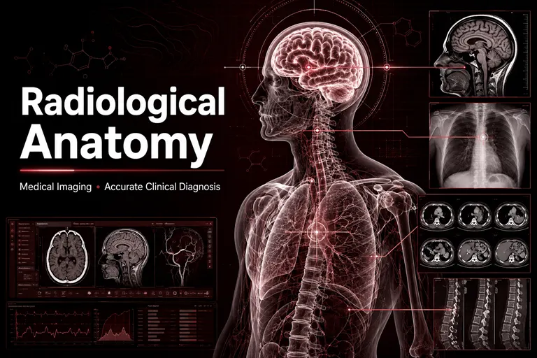
Radiological anatomy is the study of human structures as they appear across different medical imaging modalities and planes. It connects traditional anatomical knowledge with the practical requirements of diagnostic imaging. It helps clinicians understand what is normal, recognise what has changed and determine whether an unusual appearance represents disease, developmental difference or normal anatomical variation.
For the International Journal for Clinical Anatomy and Research, radiological anatomy represents an important bridge between basic anatomical science and patient care. Research in cross-sectional anatomy, anatomical variation, imaging-based morphometry and clinically applied anatomy can improve diagnostic accuracy, procedural planning and medical education.
What Is Radiological Anatomy?
Radiological anatomy, also called imaging anatomy, examines the structures of the living human body through diagnostic images. Unlike cadaveric anatomy, which involves direct observation of dissected tissues, radiological anatomy presents structures according to the physical properties detected by a particular imaging technique.
Bones, soft tissues, blood vessels, nerves and organs do not appear in the same way on every modality. Their visibility depends on factors such as tissue density, water content, blood flow, acoustic properties, imaging sequence and the use of contrast material.
Radiological anatomy therefore involves more than identifying a structure by name. It requires an understanding of:
- The normal position, size and shape of anatomical structures
- The relationships between adjacent organs and tissues
- The appearance of structures on different imaging modalities
- The effect of imaging planes and patient positioning
- Normal variations in anatomy
- Developmental and age-related differences
- Changes caused by disease, injury or surgery
- Imaging artefacts that may imitate anatomical abnormalities
A radiologist or clinician must integrate all these factors before deciding whether an image is normal or clinically significant.
The Relationship Between Anatomy and Medical Imaging
Traditional anatomy and medical imaging describe the same body from different perspectives. Dissection exposes structures directly, while imaging represents those structures through reconstructed signals or differences in tissue properties.
This distinction affects how anatomy is learned and interpreted. A medical student may first encounter the liver, brain or pelvic organs in a textbook illustration or dissection hall. In clinical practice, the same structures must be recognised as varying shades, signals and contours on a radiograph, CT scan, MRI examination or ultrasound image.
Radiological anatomy translates three-dimensional human structure into clinically meaningful visual information. It enables healthcare professionals to move between three connected forms of understanding:
- The body as a physical three-dimensional structure
- The body as represented in imaging planes
- The body as altered by disease or injury
The ability to connect these perspectives is fundamental to accurate imaging interpretation.
Understanding Anatomical Planes in Medical Imaging
Medical images are commonly displayed in axial, coronal and sagittal planes. Understanding these planes is one of the foundations of cross-sectional anatomy.
Axial Plane
The axial or transverse plane divides the body into upper and lower portions. Axial images are commonly used in CT and MRI examinations of the head, neck, chest, abdomen and pelvis.
Clinicians must learn to follow structures across a sequence of axial slices. A single image shows only one level, while the complete series reveals how a structure begins, changes shape and relates to neighbouring tissues.
Coronal Plane
The coronal plane divides the body into front and back portions. It is useful for examining structures that extend vertically, including the brain, paranasal sinuses, lungs, kidneys, spine and joints.
Coronal images may provide a more intuitive view of bilateral relationships and help demonstrate the superior and inferior extent of a lesion.
Sagittal Plane
The sagittal plane divides the body into right and left portions. It is particularly useful for assessing the brain, spinal column, pelvis, joints and midline structures.
Sagittal imaging can show curvature, alignment, depth and relationships that may be difficult to appreciate on axial images alone.
Multiplanar and Three-Dimensional Reconstruction
Modern imaging systems can reconstruct data in multiple planes or produce three-dimensional visualisations. These techniques help clinicians evaluate fractures, tumours, blood vessels, congenital abnormalities and complex surgical regions.
Nevertheless, reconstructed images should be interpreted alongside the original image data. The quality of a reconstruction depends on acquisition technique, slice thickness, software processing and image resolution.
Radiological Anatomy Across Major Imaging Modalities
Each imaging modality presents anatomy differently. Accurate interpretation requires familiarity with both the anatomical region and the technical characteristics of the examination.
Conventional Radiography
Conventional radiography uses X-rays to create two-dimensional images. It remains widely used because it is fast, accessible and particularly effective for evaluating bones, the chest and selected abdominal conditions.
Projection and Superimposition
A radiograph compresses three-dimensional anatomy into a two-dimensional image. Multiple structures may overlap, making interpretation dependent on a strong understanding of projectional anatomy.
For example, a chest radiograph includes the lungs, ribs, heart, mediastinum, diaphragm, vessels and soft tissues within one projection. A clinician must recognise their expected contours and relationships while accounting for rotation, inspiration and patient positioning.
Anatomical Landmarks on Radiographs
Landmarks help assess alignment, symmetry, organ size and possible pathology. In skeletal imaging, landmarks may indicate fractures, dislocations or degenerative changes. In chest radiography, changes in expected borders can suggest collapse, fluid, enlargement or a space-occupying lesion.
An apparent abnormality may also result from poor positioning or overlapping normal structures. Knowledge of projectional anatomy helps reduce such errors.
Computed Tomography Anatomy
Computed tomography creates detailed cross-sectional images using X-rays and computer reconstruction. It is widely used in trauma, emergency medicine, oncology, vascular assessment and the evaluation of the chest, abdomen, pelvis, brain and skeletal system.
CT can demonstrate the shape, size, density and location of internal structures while reducing the problem of overlapping anatomy seen in conventional radiography. It can also show the relationship of disease to surrounding tissues, which is valuable for diagnosis and treatment planning. RadiologyInfo.org
Tissue Density and CT Windows
Different tissues absorb X-rays to different degrees. Bone, air, fat, fluid and soft tissue therefore appear with different densities.
CT window settings can be adjusted to emphasise particular tissues. A lung window is used to examine pulmonary structures, while a bone window improves the visibility of cortical and trabecular anatomy. Soft-tissue windows assist with the evaluation of organs, muscles, vessels and pathological masses.
The ability to select and interpret appropriate windows requires knowledge of both anatomy and imaging principles.
Contrast-Enhanced CT
Contrast material can improve the visualisation of vessels, organs and abnormal tissue. The appearance of a structure may change according to the timing of image acquisition after contrast administration.
Arterial, venous and delayed phases can reveal different anatomical and pathological information. Understanding vascular anatomy and organ perfusion is therefore necessary when evaluating contrast-enhanced CT studies.
CT Angiography
CT angiography provides detailed images of arteries and veins. It can help identify aneurysms, stenosis, occlusion, vascular injury and variations in branching patterns. It also allows clinicians to assess the relationship between vessels and surrounding structures. RadiologyInfo.org
Magnetic Resonance Imaging Anatomy
Magnetic resonance imaging uses magnetic fields and radiofrequency signals to produce detailed images without ionising radiation. It is particularly valuable for examining the brain, spinal cord, joints, muscles, ligaments, pelvic organs and other soft tissues.
Tissue Contrast and MRI Sequences
MRI does not produce one fixed appearance. Different sequences emphasise different tissue characteristics. The same structure can appear bright on one sequence and dark on another.
This makes MRI highly informative but also more complex to interpret. Accurate reading requires an understanding of normal anatomy, sequence characteristics and pathological signal changes.
Neurological and Musculoskeletal Anatomy
MRI provides detailed visualisation of the brain, spinal cord, intervertebral discs, nerves, muscles, tendons, ligaments and cartilage. It helps clinicians identify subtle structural changes that may not be evident on conventional radiographs.
In neurological imaging, accurate localisation depends on knowledge of brain regions, white-matter pathways, ventricles, cranial nerves and vascular territories. In musculoskeletal imaging, interpretation requires knowledge of joint compartments, attachments and functional anatomical relationships.
Functional and Advanced MRI
Advanced techniques can provide information beyond basic structure. Diffusion imaging, perfusion studies, functional MRI and specialised sequences can assist in evaluating tissue characteristics and physiological processes.
However, these findings must still be anatomically localised. Functional information becomes clinically meaningful when it is connected to the correct organ, region or pathway.
Ultrasound Anatomy
Ultrasonography uses high-frequency sound waves to create real-time images. It is used in obstetrics, emergency medicine, cardiology, anaesthesia, vascular assessment, abdominal imaging and procedural guidance.
Dynamic and Operator-Dependent Imaging
Ultrasound differs from CT and MRI because the operator actively controls the imaging plane. Probe position, pressure, angle and orientation affect what appears on the screen.
The operator must mentally connect the image with the probe’s position on the patient. Strong knowledge of surface and regional anatomy is therefore essential.
Identifying Tissue Patterns
Fluid, bone, air and soft tissues interact differently with ultrasound waves. Their appearance depends on their acoustic properties.
Clinicians must recognise the expected patterns of organs, vessels, muscles, tendons and nerves. They must also distinguish genuine anatomical findings from shadowing, enhancement and other artefacts.
Ultrasound-Guided Procedures
Ultrasound is increasingly used to guide vascular access, nerve blocks, biopsies, drainage and fluid aspiration. Real-time imaging can improve procedural precision, but safe performance still requires knowledge of the target structure and nearby vessels, nerves and organs.
Nuclear Medicine and Hybrid Imaging
Nuclear medicine uses small quantities of radioactive tracers to evaluate physiological activity. Unlike imaging methods that primarily show structure, nuclear medicine can demonstrate metabolic or functional processes.
Hybrid techniques such as PET-CT and SPECT-CT combine functional information with anatomical localisation. A region of abnormal tracer uptake becomes more useful when it can be accurately matched to a particular organ, lymph node, bone or tissue compartment.
This combination is especially valuable in oncology, cardiology, neurology and the investigation of infection or inflammation. Diagnostic imaging plays a central role in cancer detection, staging, treatment planning, response assessment and follow-up. International Atomic Energy Agency
How Radiological Anatomy Supports Accurate Clinical Diagnosis
Medical imaging contributes to diagnosis only when findings are interpreted correctly and integrated with clinical information.
Localising Disease
Symptoms may identify a general region, but imaging can help localise the exact structure involved. A neurological deficit may originate from the brain, spinal cord, nerve root or peripheral nerve. Abdominal pain may arise from an organ, vessel, bowel segment or musculoskeletal structure.
Radiological anatomy helps narrow this localisation and supports a more focused differential diagnosis.
Defining the Extent of Abnormality
Imaging can show not only whether a lesion is present but also its size, boundaries and relationship to surrounding tissues. This information is important when assessing tumours, infections, trauma and inflammatory disease.
For example, determining whether a mass is confined to one compartment or has crossed a fascial plane can influence staging and treatment planning.
Differentiating Similar Conditions
Different diseases can produce similar symptoms. Imaging features, anatomical location and patterns of spread can help distinguish among them.
An accurate diagnosis may depend on recognising:
- The organ from which a mass originates
- The vascular territory affected by an infarction
- The joint compartment containing an injury
- The distribution of infection
- The pathway followed by fluid or blood
- Whether a structure is displaced, compressed or invaded
These distinctions require detailed anatomical reasoning.
Correlating Imaging with Clinical Findings
A scan should not be interpreted in isolation. The importance of an imaging finding depends on whether it explains the patient’s symptoms and examination results.
Some abnormalities may be incidental. Others may appear subtle but have major clinical significance. Radiological anatomy helps clinicians assess whether the location and relationships of a finding are consistent with the clinical presentation.
Anatomical Variations in Diagnostic Imaging
Anatomical variation is a major consideration in radiology. The human body does not follow one perfectly uniform pattern. Variations occur in vessels, ducts, bones, muscles, nerves and organs.
Variations That Resemble Disease
Some variations can imitate tumours, fractures, vascular abnormalities or other pathological findings. Recognising normal variants can prevent unnecessary investigations, treatment or patient anxiety.
Examples may include unusual vessel courses, accessory bones, variant muscles, altered organ positions and developmental remnants.
Clinically Significant Variations
A variation may be normal but still affect clinical care. Vascular branching patterns can influence surgery and interventional procedures. Variation in biliary, renal or pulmonary anatomy can alter operative planning. Differences in spinal anatomy may affect level identification or needle placement.
The significance of a variant therefore depends on context. The radiologist should describe it when it may influence diagnosis, treatment or procedural safety.
Age-Related and Developmental Anatomy
Anatomy changes throughout life. Paediatric imaging requires awareness of normal growth, incomplete skeletal maturation and developmental differences. These appearances should not be mistaken for injury or disease.
Ageing also produces structural changes that may be expected rather than pathological. The interpreter must distinguish normal age-related findings from clinically important abnormalities.
Radiological Anatomy in Surgical and Treatment Planning
Medical imaging allows surgical teams to understand a patient’s individual anatomy before treatment. This is particularly valuable when anatomy has been altered by disease, trauma, congenital difference or previous surgery.
Preoperative Assessment
Preoperative imaging can demonstrate:
- The precise location and extent of a lesion
- Its relationship to vessels, nerves and organs
- Possible routes of surgical access
- Anatomical variations
- Tissue invasion or displacement
- Previous operative changes
- Potential complications
This information can help surgeons select an appropriate approach and prepare for structures that may be encountered.
Minimally Invasive and Image-Guided Treatment
Interventional radiology, endovascular procedures, endoscopy and minimally invasive surgery require accurate anatomical navigation. Instruments may be guided through vessels, ducts, cavities or narrow tissue pathways.
Imaging and anatomical knowledge work together to improve precision while limiting injury to surrounding structures.
Radiotherapy Planning
Radiotherapy requires accurate localisation of the target while protecting nearby healthy tissues. Cross-sectional anatomy helps teams define treatment areas and recognise organs at risk.
Small errors in anatomical identification may affect treatment effectiveness or increase toxicity. Careful imaging interpretation and multidisciplinary review are therefore essential.
Radiological Anatomy and Patient Safety
Radiological anatomy contributes to patient safety at several stages of care.
Preventing Diagnostic Errors
Misidentifying a normal structure as disease can lead to unnecessary procedures. Failing to recognise an abnormality can delay treatment. Errors may also occur when a lesion is assigned to the wrong organ or anatomical compartment.
Systematic image review, appropriate clinical information and strong anatomical training can reduce these risks.
Avoiding Procedural Injury
Image guidance can help clinicians see the target and nearby structures during biopsy, drainage, vascular access or injection. However, safe intervention still depends on understanding the anatomy beyond the immediate needle or catheter path.
Appropriate Imaging Selection
Different clinical questions require different imaging modalities. The best examination is not always the one that provides the greatest amount of detail. The choice should consider the suspected condition, anatomical region, urgency, patient characteristics and potential risks.
Radiation Protection
CT, radiography, fluoroscopy and nuclear medicine involve ionising radiation. Examinations should be justified and performed using protocols that provide sufficient diagnostic information while avoiding unnecessary exposure.
Radiation optimisation is particularly important for children, pregnant patients and individuals requiring repeated imaging. The International Atomic Energy Agency provides guidance on patient-dose assessment, quality assurance and optimisation across diagnostic radiology. IAEA radiation-protection resources
The Role of Radiological Anatomy in Medical Education
Radiological anatomy helps students connect foundational science with living patients. Integrating imaging into anatomy education can improve spatial understanding and demonstrate why anatomical relationships matter in clinical practice.
Connecting Dissection with Living Anatomy
Cadaveric dissection allows direct study of structures, while imaging shows how those structures appear in living individuals. Used together, these methods provide complementary perspectives.
Students can compare a dissected region with CT or MRI images and learn how organs, tissue planes and anatomical spaces translate into clinical imaging.
Developing Three-Dimensional Understanding
A sequence of cross-sectional images requires the learner to reconstruct anatomy mentally. This process strengthens spatial reasoning, which is important in radiology, surgery, emergency medicine and procedural specialties.
Case-Based Learning
Imaging cases can connect anatomy with symptoms, examination findings and pathology. Instead of learning structures as isolated facts, students understand how changes in those structures affect patients.
Training Beyond Radiology
Radiological anatomy is important for surgeons, physicians, anaesthesiologists, emergency clinicians, physiotherapists and other healthcare professionals. Point-of-care ultrasonography and image-guided procedures have expanded the need for imaging literacy across disciplines.
Research Opportunities in Radiological Anatomy
Radiological anatomy continues to offer valuable research opportunities. Imaging enables researchers to study living anatomy, quantify variation and examine relationships across different populations.
Relevant areas include:
- Cross-sectional anatomical measurements
- Vascular variations
- Organ morphology and volume
- Radiological assessment of developmental anatomy
- Age-related anatomical changes
- Three-dimensional reconstruction
- Imaging-based surgical planning
- Comparison of cadaveric and radiological findings
- Artificial intelligence for anatomical segmentation
- Digital anatomy education
- Interobserver reliability in anatomical measurement
- Population-specific morphometric reference data
High-quality studies should clearly explain their clinical relevance. A measurement becomes more meaningful when researchers show how it may improve diagnosis, surgical planning, implant selection, procedural safety or interpretation of variation.
Researchers must also report imaging protocols, patient-selection criteria, measurement techniques, ethical approval, observer reliability and statistical methods transparently.
Artificial Intelligence and the Future of Imaging Anatomy
Artificial intelligence is increasingly used in medical imaging for reconstruction, segmentation, detection, measurement and workflow prioritisation.
Automated Anatomical Segmentation
AI systems can identify and outline organs, tissues or lesions. This may support treatment planning, volumetric analysis and research. Automated segmentation can reduce repetitive work, but its accuracy should be reviewed carefully, particularly in patients with unusual anatomy or extensive disease.
Computer-Assisted Detection
AI can highlight areas that may require attention. These systems may support radiologists, but they do not remove the need for professional interpretation.
A flagged region must still be anatomically localised and assessed in the context of the entire examination and the patient’s clinical information.
Limitations of AI in Anatomical Interpretation
AI models are influenced by their training data. If certain populations, uncommon variations or rare diseases are underrepresented, performance may be less reliable in those cases.
Human expertise remains necessary to identify unexpected relationships, assess image quality, recognise artefacts and determine whether a finding is clinically meaningful.
Patient-Specific Imaging and Precision Care
Future imaging systems will increasingly combine anatomy, function and patient history. Three-dimensional models and augmented-reality tools may assist surgery and intervention by displaying patient-specific structures.
These developments will increase, rather than reduce, the importance of accurate radiological anatomy.
The Role of IJCAR in Advancing Radiological Anatomy
The International Journal for Clinical Anatomy and Research provides a relevant scholarly platform for work connecting human anatomy with diagnostic imaging and clinical care.
Within its focus on clinical, gross, developmental, histological and radiological anatomy, IJCAR can support research that:
- Identifies clinically significant anatomical variations
- Establishes imaging-based morphometric reference data
- Connects cadaveric findings with living anatomy
- Improves surgical and procedural planning
- Evaluates methods of radiological anatomy education
- Examines anatomical changes associated with age or development
- Applies three-dimensional imaging to clinical problems
- Strengthens collaboration among anatomists, radiologists and surgeons
Original studies, systematic reviews, educational research and carefully documented anatomical case reports can all contribute to this field when they are methodologically sound and clinically relevant.
Authors interested in submitting work should review the journal’s aims and scope, ethical policies and manuscript instructions before submission.
Frequently Asked Questions
What is radiological anatomy?
Radiological anatomy is the study of human structures as they appear on X-rays, CT scans, MRI examinations, ultrasound and other imaging modalities. It connects anatomical knowledge with clinical image interpretation.
Why is radiological anatomy important for diagnosis?
It helps clinicians identify normal structures, recognise abnormalities, localise disease and distinguish pathological findings from normal anatomical variations.
What is cross-sectional anatomy?
Cross-sectional anatomy examines the body in slices or planes, usually axial, coronal and sagittal. It is essential for interpreting CT and MRI examinations.
How does CT differ from MRI in anatomical imaging?
CT uses X-rays and is particularly effective for evaluating bone, trauma, lungs and many emergency conditions. MRI uses magnetic fields and provides detailed soft-tissue contrast, especially for the brain, spinal cord, joints and muscles. The appropriate modality depends on the clinical question.
Why can anatomical variations cause diagnostic errors?
Some variations resemble disease, while others change expected structural relationships. If the interpreter is unfamiliar with a variant, it may be incorrectly reported as an abnormality or overlooked during treatment planning.
How does radiological anatomy improve surgical safety?
It helps surgeons identify the position of lesions, nearby vessels and nerves, possible anatomical variations and safe routes of access before an operation.
Is radiological anatomy relevant outside radiology?
Yes. It is important in surgery, emergency medicine, anaesthesia, orthopaedics, neurology, oncology, physiotherapy and any field involving imaging or image-guided procedures.
Can artificial intelligence replace anatomical knowledge in radiology?
No. AI can assist with segmentation, measurement and detection, but its output must be reviewed by trained professionals. Anatomical expertise remains necessary for interpreting variations, artefacts and complex clinical relationships.
What radiological anatomy research can be submitted to IJCAR?
Potential topics include imaging-based morphometry, anatomical variations, cross-sectional anatomy, three-dimensional reconstruction, developmental imaging, radiological case reports and studies connecting imaging findings with surgical or clinical practice. Authors should confirm compatibility with the journal’s latest submission guidelines.
Conclusion
Radiological anatomy is the language through which medical images become clinically meaningful. It enables healthcare professionals to identify normal structures, recognise disease, interpret anatomical variations and connect imaging findings with a patient’s symptoms and treatment needs.
Its importance extends beyond diagnostic radiology. Surgeons use imaging anatomy to plan operations, clinicians use it to localise disease, interventional specialists rely on it to navigate safely and medical educators use it to connect foundational anatomy with living patients.
As imaging technologies become more detailed and artificial intelligence becomes increasingly integrated into radiology, anatomical expertise will remain indispensable. Technology can improve visualisation and support detection, but accurate diagnosis still requires clinical reasoning, anatomical knowledge and responsible professional judgement.
Through research in clinical and radiological anatomy, the International Journal for Clinical Anatomy and Research can help strengthen the connection between structural science, diagnostic accuracy and patient safety. Researchers working in imaging-based anatomy, anatomical variation, morphometry and clinically applied imaging are encouraged to consult IJCAR’s submission guidelines and contribute relevant scholarly work.
For medical institutions, scholarly societies or journal founders planning a professional research journal, support is available for ScholarJMS website and editorial workflow setup, OJSCloud hosting and migration, ISSN consulting, and GetDOI Crossref DOI workflow guidance. Contact +91 82003 85143 via WhatsApp or email inquiry@ojscloud.com.