Clinical Anatomy in Modern Medicine: Its Role in Diagnosis, Surgery and Patient Safety
Clinical anatomy is the practical application of anatomical knowledge to the diagnosis, treatment and prevention of disease. It connects the structural organisation of the human body with the decisions healthcare professionals make during physical examination, medical imaging, surgery, emergency care and other clinical procedures.
Although modern medicine increasingly relies on advanced imaging, minimally invasive surgery, robotics and artificial intelligence, these technologies have not reduced the importance of anatomy. They have made precise anatomical knowledge even more valuable. A radiological image must be interpreted in relation to normal structures. A surgical instrument must be guided through a safe anatomical pathway. A clinical symptom must be connected to the organ, nerve, vessel, tissue or region that may be involved.
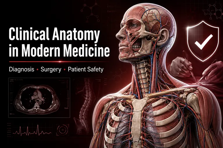
Clinical anatomy therefore remains one of the foundations of safe and effective medical practice. It enables clinicians to translate what they observe on the body surface, in an imaging study or during an operation into an informed understanding of the structures beneath.
For the International Journal for Clinical Anatomy and Research, this relationship between anatomical science and clinical practice represents an important field of scholarly enquiry. Research involving gross anatomy, clinical anatomy, developmental anatomy, histology, radiological anatomy and anatomical variations can directly improve clinical reasoning, procedural safety and patient outcomes.
What Is Clinical Anatomy?
Clinical anatomy, sometimes described as applied anatomy, examines the structures of the human body in relation to healthcare practice. Traditional anatomy explains where structures are located, how they are organised and how they relate to one another. Clinical anatomy extends that knowledge by asking how these relationships affect diagnosis, disease, surgery and treatment.
For example, knowing the course of a peripheral nerve is a matter of descriptive anatomy. Understanding how that nerve may be compressed, injured during surgery or examined in a patient is clinical anatomy. Similarly, identifying the normal branches of an artery is anatomical knowledge, while recognising how variations in those branches could affect a vascular procedure is applied clinical knowledge.
Clinical anatomy encompasses several connected areas:
- Gross and regional anatomy
- Surface and living anatomy
- Surgical anatomy
- Radiological and cross-sectional anatomy
- Neuroanatomy
- Developmental anatomy and embryology
- Histological anatomy
- Anatomical variation
- Functional and biomechanical anatomy
These areas are not isolated. In clinical practice, they frequently overlap. A surgeon planning a procedure may need regional, radiological and vascular anatomy simultaneously. A clinician assessing a neurological deficit may combine surface anatomy, neuroanatomy and imaging findings to identify the likely site of injury.
Why Clinical Anatomy Remains Essential in Modern Medicine
Medical technology can display the body in increasingly sophisticated ways, but technology does not remove the need for anatomical interpretation. Computed tomography, magnetic resonance imaging, ultrasonography, endoscopy, robotic surgery and image-guided procedures all depend on the user’s ability to recognise structures and understand their spatial relationships.
Clinical anatomy provides the framework through which medical information becomes meaningful. It helps healthcare professionals determine:
- Which structure may be producing a patient’s symptoms
- How a disease has altered normal anatomy
- Whether an image demonstrates normal anatomy or pathology
- Which surgical or procedural route is safest
- What anatomical variations may be encountered
- Which nearby structures could be injured during treatment
- How complications may be prevented or managed
Anatomical understanding is therefore not simply a subject learned during the early years of medical education. It is a continuing clinical competency that must be reinforced as diagnostic and therapeutic practices evolve.
The Role of Clinical Anatomy in Medical Diagnosis
Accurate diagnosis begins with understanding where symptoms originate. Pain, weakness, swelling, sensory loss and restricted movement often follow recognisable anatomical distributions. Clinical anatomy allows healthcare professionals to connect these findings with the structures most likely to be involved.
Physical Examination and Surface Anatomy
Surface anatomy helps clinicians identify internal structures through visible or palpable external landmarks. These landmarks guide routine examination and assist with the assessment of organs, bones, muscles, vessels and nerves.
The location of a pulse, the boundaries of an organ, the movement of a joint and the distribution of cutaneous sensation all depend on anatomical relationships. Accurate examination requires more than memorising a series of steps. The clinician must understand what lies beneath the surface and why a particular finding may be significant.
Surface landmarks also guide procedures such as:
- Intramuscular injections
- Venous and arterial access
- Lumbar puncture
- Chest drainage
- Joint aspiration
- Regional anaesthesia
- Biopsy and tissue sampling
An inaccurate understanding of these landmarks can lead to failed procedures, delayed diagnosis or avoidable injury.
Localising Neurological Lesions
Clinical neuroanatomy is especially important because neurological symptoms frequently reveal the probable location of a lesion. Patterns of motor weakness, sensory loss, altered reflexes or cranial nerve dysfunction can help determine whether the abnormality is located in the brain, spinal cord, nerve root, plexus or peripheral nerve.
The distribution of symptoms is rarely random. It often follows established anatomical pathways. By combining examination findings with neuroanatomical knowledge, clinicians can narrow the differential diagnosis and select appropriate imaging or laboratory investigations.
Understanding Referred Pain
Pain may be experienced in a location different from the diseased organ or tissue. Referred pain occurs because sensory information from different structures may converge on common neural pathways.
A sound understanding of segmental innervation and visceral anatomy helps clinicians interpret these patterns. Without this knowledge, pain originating from an internal organ could be mistaken for a musculoskeletal condition or another unrelated disorder.
Distinguishing Normal Anatomy from Disease
Not every unusual finding represents pathology. Some findings are normal anatomical variations. Others may reflect age, sex, body composition, developmental history or previous treatment.
Clinical anatomy helps clinicians recognise the boundaries between normality, variation and disease. This distinction can prevent unnecessary investigations and reduce the risk of misdiagnosis.
Clinical Anatomy and Medical Imaging
Radiological anatomy has become one of the most important bridges between anatomical science and clinical care. Modern imaging allows clinicians to examine the living body in two-dimensional, three-dimensional and dynamic formats.
Cross-Sectional Anatomy
Computed tomography and magnetic resonance imaging present the body in axial, coronal, sagittal and reconstructed planes. These images require a clear understanding of cross-sectional anatomy.
Clinicians must identify normal tissues, recognise anatomical boundaries and assess spatial relationships. A mass may compress a vessel, displace an organ or invade an adjacent tissue plane. The significance of such findings can only be understood through anatomical interpretation.
Ultrasonography
Ultrasonography is widely used in obstetrics, emergency medicine, cardiology, anaesthesia, surgery and image-guided intervention. It provides real-time visualisation but can be technically dependent on the operator.
Knowledge of regional anatomy helps the operator position the probe, interpret tissue patterns and avoid confusing one structure with another. It is particularly important when ultrasound is used to guide vascular access, nerve blocks, fluid aspiration or biopsy.
Interventional Radiology
Interventional radiology uses imaging guidance to perform diagnostic and therapeutic procedures through small access points. Catheters, wires and needles must pass through carefully selected anatomical routes.
Successful intervention requires knowledge of vascular territories, organ relationships, tissue planes and potential variants. Detailed anatomical planning can help shorten procedure time, improve precision and reduce complications.
Three-Dimensional Imaging and Digital Reconstruction
Three-dimensional reconstructions can improve the visualisation of fractures, congenital abnormalities, tumours, vascular networks and complex surgical regions. They can also support patient-specific planning.
However, a digital model is only as useful as its clinical interpretation. Anatomical expertise remains necessary to determine which relationships are important and how they affect the intended procedure.
The Role of Clinical Anatomy in Surgery
Surgery is one of the clearest demonstrations of applied anatomy. Every operation involves movement through anatomical layers and around structures that must be preserved, repaired, removed or reconstructed.
Surgical Planning
Preoperative planning includes understanding the location of the target lesion, the safest route of access and the structures that may be encountered. The approach must provide adequate exposure while limiting damage to healthy tissue.
Surgeons consider:
- The position and depth of the target structure
- Nearby arteries, veins and nerves
- Fascial planes and tissue compartments
- Possible anatomical variations
- Previous surgery, trauma or disease-related distortion
- The availability of alternative operative routes
The quality of this planning can influence operative time, blood loss, postoperative function and recovery.
Identifying Safe Surgical Planes
Natural tissue planes can provide relatively safe pathways during surgery. Understanding fascial layers, spaces and anatomical compartments helps surgeons separate tissues with less trauma.
If the correct plane is missed, dissection may become more difficult and the risk of bleeding, nerve injury or organ damage may increase. Clinical anatomy research can refine the description of these planes and help improve operative techniques.
Minimally Invasive and Robotic Surgery
Laparoscopic, endoscopic and robotic techniques offer smaller incisions and potentially faster recovery, but they also change the surgeon’s visual and tactile experience. Structures may appear from unfamiliar angles, and direct palpation may be limited.
These procedures require a strong three-dimensional mental model of the operative region. Surgeons must interpret magnified images, control instruments remotely and anticipate structures outside the visible field.
As minimally invasive approaches expand, the relationship between anatomy, imaging and technology becomes increasingly important.
Reconstructive and Orthopaedic Surgery
Reconstruction requires more than removing abnormal tissue. It may involve restoring form, movement, stability, vascular supply or nerve function.
Orthopaedic and reconstructive procedures depend on detailed knowledge of bones, joints, muscles, tendons, ligaments, nerves and vascular pedicles. Research on the anatomical characteristics of these structures can influence graft selection, implant placement and rehabilitation planning.
Anatomical Variations and Their Clinical Importance
Human anatomy is not identical in every individual. Variations may occur in arteries, veins, nerves, muscles, bones, organs and developmental structures. Many are harmless and remain undetected throughout life, but some become clinically important during diagnosis or treatment.
Variations in Surgery
An unexpected artery or an unusual nerve pathway may increase the risk of bleeding or neurological injury. Awareness of frequently reported variations helps surgeons anticipate these possibilities.
Preoperative imaging may reveal some variants, but not all are identified before an operation. Surgeons must therefore combine preparation with the ability to recognise unfamiliar anatomy during a procedure.
Variations in Radiology
An anatomical variant may resemble a mass, obstruction, vascular abnormality or other pathological process. Radiologists must be familiar with common and uncommon variants so that normal differences are not incorrectly classified as disease.
At the same time, a variation should not automatically be dismissed as incidental. Its clinical significance depends on its location, relationship to symptoms and relevance to planned treatment.
Variations in Medical Procedures
The success of catheterisation, nerve blocks, injections and other procedures may be affected by differences in anatomy. A standard technique may require modification when usual landmarks are absent or when a structure follows an atypical course.
Carefully documented cadaveric studies, imaging research and clinical case reports help build the evidence clinicians need to manage these situations.
Clinical Anatomy as a Foundation of Patient Safety
Patient safety depends on recognising risk before harm occurs. Anatomical knowledge contributes to this objective by helping healthcare professionals identify vulnerable structures and select safer techniques.
Preventing Iatrogenic Injury
Iatrogenic injuries are complications unintentionally caused during medical care. Some arise when a nerve, blood vessel, duct or organ lies close to a procedural pathway.
Knowledge of anatomical danger zones can help clinicians modify incision placement, needle direction, dissection technique or instrument depth. Clear reporting of anatomy-related complications also allows the wider medical community to learn from difficult cases.
Safer Injections and Vascular Access
Even common procedures require anatomical precision. An injection administered at an inappropriate site may injure a nerve or vessel. Incorrect vascular access can lead to bleeding, thrombosis, pneumothorax or damage to adjacent structures.
Anatomical training, ultrasound guidance and standardised procedural protocols work together to reduce these risks.
Reducing Wrong-Site and Wrong-Level Procedures
Precise anatomical localisation is necessary to ensure that treatment is delivered to the correct side, level or structure. This is particularly important in spinal procedures, limb surgery, neurosurgery and image-guided intervention.
Safety checklists are valuable, but they must be supported by accurate clinical examination, imaging correlation and anatomical identification.
Recognising Complications Early
Anatomical knowledge also helps clinicians understand how complications may spread or present. Blood, infection, air or fluid may travel through predictable spaces and tissue planes. Recognising these pathways can support earlier diagnosis and treatment.
Clinical Anatomy in Medical Education and Training
Anatomy education is changing as medical curricula adopt integrated, competency-based and clinically oriented approaches. Cadaveric dissection continues to provide valuable spatial understanding, but it is now often combined with imaging, digital models, simulation, prosection, virtual reality and case-based learning.
Cadaveric Dissection
Cadaveric study offers direct exposure to anatomical depth, texture, variation and spatial relationships. It also introduces students to professional conduct, respect for donors and ethical responsibility.
For surgical trainees and researchers, cadaveric work can provide an environment for examining approaches, testing devices and studying variations before applying new techniques in patient care.
Simulation and Virtual Anatomy
Simulation enables learners to practise procedures without placing patients at risk. Virtual and augmented reality can provide repeatable visualisation of complex anatomical regions.
These tools are most effective when they supplement sound anatomical teaching. Digital models may simplify structures and cannot always reproduce the variability seen in real patients or cadaveric material.
Clinically Integrated Learning
Anatomy becomes more memorable when students understand its clinical relevance. Linking anatomical regions with imaging, physical examination, pathology and procedural cases encourages learners to apply knowledge rather than memorise isolated facts.
This approach can support better long-term retention and prepare students for clinical decision-making.
The Contribution of Clinical Anatomy Research
Clinical anatomy is not a static body of established facts. Research continues to refine anatomical descriptions, quantify variations and evaluate their clinical importance.
Important research approaches include:
- Cadaveric dissection and morphometric analysis
- Histological and histomorphometric investigation
- Radiological measurement and image-based analysis
- Surgical and procedural observations
- Anatomical case reports
- Developmental and embryological studies
- Systematic reviews and evidence synthesis
- Three-dimensional modelling
- Biomechanical investigation
- Digital and technology-assisted anatomy education
Well-designed studies should explain why an anatomical observation matters clinically. Measuring a structure is useful, but the research becomes more valuable when it clarifies how the measurement may affect diagnosis, implant selection, surgical access, imaging interpretation or patient safety.
Researchers should also report methodology transparently. Sample characteristics, measurement techniques, imaging protocols, observer reliability, ethical considerations and study limitations all influence whether findings can be applied responsibly.
The Role of IJCAR in Advancing Applied Anatomical Knowledge
The International Journal for Clinical Anatomy and Research provides a scholarly platform for research across human anatomy and its clinical applications. Its scope includes clinical, gross, developmental, histological and radiological anatomy, enabling researchers to connect foundational anatomical investigation with real medical needs.
The journal can contribute to the discipline by encouraging research that:
- Documents clinically relevant anatomical variations
- Improves understanding of surgical approaches
- Connects radiological findings with structural anatomy
- Evaluates anatomy education and training methods
- Examines developmental and histological relationships
- Supports safer clinical procedures
- Presents unusual cases with clear educational value
- Promotes interdisciplinary collaboration between anatomists and clinicians
Original research, systematic reviews, case reports and short communications can each make a valuable contribution when they are scientifically rigorous and clinically meaningful.
Researchers interested in these areas may review IJCAR’s aims and scope, author instructions and manuscript-submission requirements before preparing their work.
Future Directions in Clinical Anatomy
Clinical anatomy will continue to develop alongside precision medicine, artificial intelligence, three-dimensional printing and personalised surgery.
Artificial Intelligence and Anatomical Imaging
Artificial intelligence can assist with image segmentation, structural recognition and measurement. It may help identify anatomical landmarks or highlight abnormal findings. Nevertheless, clinical oversight remains essential because algorithms can be affected by image quality, training data and unusual anatomy.
Research should evaluate not only whether an algorithm performs accurately, but also how it responds to anatomical variations and complex clinical cases.
Patient-Specific Anatomical Planning
Three-dimensional models based on individual imaging data can help clinicians understand complex anatomy before treatment. Patient-specific planning may be especially valuable in craniofacial surgery, orthopaedics, neurosurgery, cardiovascular intervention and tumour resection.
Three-Dimensional Printing
Printed anatomical models can support education, procedural rehearsal and patient communication. They allow clinicians to examine relationships physically and may be useful when conventional images are difficult to interpret.
Image-Guided and Robotic Procedures
Navigation systems and robotic platforms are likely to make procedures more precise, but safe use will still depend on anatomical registration and recognition. Errors in localisation or model alignment can have serious consequences.
Interdisciplinary Collaboration
Future progress will require collaboration among anatomists, surgeons, radiologists, pathologists, medical educators, biomedical engineers and data scientists. Such partnerships can transform anatomical observations into practical improvements in diagnosis and treatment.
Frequently Asked Questions
What is the main purpose of clinical anatomy?
The main purpose of clinical anatomy is to apply knowledge of human structure to medical practice. It helps clinicians understand symptoms, interpret diagnostic images, plan procedures, perform surgery and reduce avoidable injury.
How does clinical anatomy support accurate diagnosis?
It enables healthcare professionals to connect symptoms and examination findings with specific organs, nerves, vessels, muscles and anatomical regions. It also helps distinguish normal variations from disease.
Why is anatomy important when modern imaging is available?
Imaging shows structures, but anatomical knowledge is required to identify and interpret them. CT, MRI, ultrasound and other technologies cannot be used effectively without understanding normal relationships and possible variations.
What is the difference between clinical anatomy and surgical anatomy?
Clinical anatomy covers the broader application of anatomy to diagnosis, examination, imaging, treatment and procedures. Surgical anatomy focuses particularly on the structures and relationships relevant to operative access and surgical safety.
Why are anatomical variations important?
Variations can affect diagnosis, imaging interpretation, surgical planning and procedural success. An unexpected vascular or neural pattern may increase the risk of complications if it is not recognised.
How does clinical anatomy improve patient safety?
It helps clinicians identify safe procedural routes, protect nearby structures, recognise danger zones, prevent wrong-site interventions and anticipate complications based on anatomical pathways.
Is cadaveric research still relevant?
Yes. Cadaveric research remains valuable for studying spatial relationships, surgical approaches, morphometry and anatomical variations. It also supports procedural training and the development of new techniques.
What types of clinical anatomy research can be submitted to IJCAR?
Relevant work may include original anatomical research, cadaveric studies, morphometric investigations, radiological anatomy, histological research, anatomical case reports, systematic reviews, developmental anatomy and clinically relevant educational studies. Authors should confirm alignment with the journal’s current aims, scope and submission guidelines.
Conclusion
Clinical anatomy remains indispensable to modern medicine. It supports accurate physical examination, diagnostic reasoning, imaging interpretation, surgical planning and the safe performance of medical procedures. Its importance is increasing as healthcare becomes more technologically advanced, image-guided and patient-specific.
The central lesson is clear: technology can enhance anatomical vision, but it cannot replace anatomical understanding. Clinicians still need to recognise normal relationships, anticipate variations and interpret structural changes in the context of an individual patient.
Through the publication of research in clinical, gross, developmental, histological and radiological anatomy, the International Journal for Clinical Anatomy and Research can help connect anatomical science with safer and more effective healthcare. Researchers working in these fields are encouraged to consult the journal’s author guidelines and consider IJCAR as a platform for sharing scientifically rigorous and clinically meaningful findings.
Journal publishers, medical institutions or scholarly societies seeking support with a professional journal website, manuscript submission system, editorial workflow, OJS migration, ISSN readiness or Crossref DOI planning may explore ScholarJMS, OJSCloud and GetDOI. For publishing guidance, contact +91 82003 85143 on WhatsApp or email inquiry@ojscloud.com.